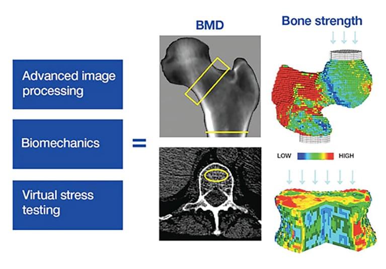
双能X线吸收法(DXA)是骨质疏松诊断和骨矿物质密度(BMD)测量的核心技术,但骨折风险并不完全由BMD决定。骨微结构、皮质骨孔隙度、骨几何形态、骨材料特性、既往骨折及跌倒风险均可能影响骨骼的力学耐受能力。随着影像学、计算生物力学和临床预测模型的发展,骨折风险评估正从单一BMD测量走向多维骨强度评价。本文探讨DXA联合骨小梁评分(TBS)、定量CT(QCT)、高分辨率外周定量CT(HR-pQCT)、有限元分析及临床风险算法的应用价值、证据边界与未来研究方向。
Dual-energy X-ray absorptiometry (DXA) remains central to osteoporosis diagnosis and bone mineral density (BMD) measurement, yet fracture risk is not determined by BMD alone. Bone microarchitecture, cortical porosity, skeletal geometry, material properties, prior fractures, and falls all influence the mechanical competence of the skeleton. Advances in imaging, computational biomechanics, and clinical prediction models are shifting fracture-risk assessment toward multidimensional evaluation of bone strength. This article examines the clinical value, evidentiary limitations, and future directions of combining DXA with trabecular bone score (TBS), quantitative CT (QCT), high-resolution peripheral quantitative CT (HR-pQCT), finite element analysis, and clinical risk algorithms.
一、为什么需要超越BMD?
DXA通过二维投影测量面积骨密度,是目前临床诊断骨质疏松和监测骨密度变化的重要标准方法。然而,面积BMD无法完整反映三维骨结构,也不能直接测量骨组织的材料强度。部分患者即使BMD尚未达到骨质疏松诊断阈值,仍可能发生脆性骨折;而部分BMD较低的患者,其实际骨折风险也会因年龄、既往骨折和其他临床因素而显著不同。
DXA estimates areal BMD from a two-dimensional projection and remains a key method for diagnosing osteoporosis and monitoring changes in bone density. However, areal BMD cannot fully characterize three-dimensional bone architecture or directly measure the material strength of bone tissue. Some patients sustain fragility fractures despite BMD values above the osteoporosis diagnostic threshold, while fracture risk among individuals with low BMD varies substantially according to age, previous fractures, and other clinical factors.
因此,未来评估的核心不应只是寻找另一种骨密度检测方法,而是整合骨量、骨结构、力学性能和个体临床风险,形成更接近骨骼真实抗骨折能力的评估框架。需要强调的是,“多维评价”并不意味着所有检查均需常规实施,而是根据患者的临床问题选择能够提供增量信息的指标。
The goal is therefore not simply to find another method of measuring bone density, but to integrate bone quantity, architecture, mechanical performance, and individual clinical risk into a framework that better reflects resistance to fracture. Multidimensional assessment does not mean that every test should be performed routinely; rather, additional measurements should be selected when they provide clinically meaningful information beyond standard assessment.
二、DXA衍生指标:在现有检查基础上增加信息
骨小梁评分(trabecular bone score,TBS)是从腰椎DXA图像的灰度纹理中提取的指标,可间接反映与骨小梁微结构相关的特征。与BMD联合使用时,TBS可为部分患者提供额外的骨折风险信息,并用于调整FRAX计算的骨折概率。国际临床骨密度测量学会(ISCD)2023年官方立场支持在适当人群中结合BMD和TBS进行风险评估,但不建议单独依据TBS决定治疗。
Trabecular bone score (TBS) is derived from grayscale texture analysis of lumbar spine DXA images and indirectly reflects features associated with trabecular microarchitecture. When combined with BMD, TBS can provide additional fracture-risk information in selected patients and can be used to adjust FRAX-derived fracture probabilities. The 2023 ISCD Official Positions support combined BMD and TBS assessment in appropriate populations, but do not recommend using TBS alone to determine treatment.
椎体骨折评估(vertebral fracture assessment,VFA)则利用DXA设备获取的侧位脊柱图像识别椎体压缩性骨折。其临床意义在于发现既往未被识别的椎体骨折,因为骨折史本身会显著影响后续骨折风险和治疗决策。对于符合适应证的患者,将VFA纳入骨密度检查流程,可能比单纯增加BMD测量次数更有临床价值。
Vertebral fracture assessment (VFA) uses lateral spine images acquired with DXA equipment to identify vertebral fractures. Its clinical value lies in detecting previously unrecognized vertebral fractures, which can substantially influence subsequent fracture risk and treatment decisions. For appropriately selected patients, incorporating VFA into the bone assessment pathway may be more clinically informative than simply repeating BMD measurements.
三、三维骨结构与力学分析:QCT、HR-pQCT和有限元分析
定量CT(quantitative computed tomography,QCT)能够测量体积骨密度(vBMD),并在一定程度上区分皮质骨与松质骨。与DXA不同,QCT可提供三维空间信息,有助于分析骨量在不同骨区的分布。但其应用需要考虑辐射剂量、设备校准、分析方法及不同平台之间的可比性。
Quantitative CT (QCT) measures volumetric BMD and can, to some extent, distinguish cortical from trabecular bone. Unlike DXA, it provides three-dimensional spatial information that supports analysis of bone distribution across different compartments. Its use nevertheless requires attention to radiation dose, calibration, analytical methods, and comparability across platforms.
高分辨率外周定量CT(HR-pQCT)可对桡骨远端和胫骨远端进行三维微结构成像,评估皮质骨厚度、皮质骨孔隙度、骨小梁数量及厚度等参数。研究显示,HR-pQCT衍生指标与脆性骨折相关,其中皮质骨体积骨密度、骨小梁厚度及估算刚度等参数具有预测潜力。不过,现有证据在不同人群中的代表性仍有限,尤其需要加强亚洲人群的验证。
High-resolution peripheral quantitative CT (HR-pQCT) provides three-dimensional microstructural imaging of the distal radius and tibia. It can quantify cortical thickness, cortical porosity, trabecular number, and trabecular thickness. Studies have linked HR-pQCT-derived parameters to fragility fractures, with cortical volumetric BMD, trabecular thickness, and estimated stiffness showing predictive potential. However, the representativeness of existing evidence across populations remains limited, and further validation in Asian populations is particularly important.


有限元分析(finite element analysis,FEA)可将CT或HR-pQCT图像转化为计算模型,在设定载荷和材料参数后估算骨骼的刚度、应力分布及失效载荷。其优势在于从结构图像进一步推断力学性能,而不是仅以骨密度作为骨强度的替代指标。但模型结果依赖图像分辨率、材料属性假设、边界条件及验证方式,不能直接等同于真实人体骨折试验。
Finite element analysis (FEA) converts CT or HR-pQCT images into computational models to estimate skeletal stiffness, stress distribution, and failure load under specified loading and material assumptions. Its principal advantage is the ability to infer mechanical performance from structural images rather than relying on BMD as a proxy for strength. However, results depend on image resolution, assumptions about material properties, boundary conditions, and validation methods; they cannot be equated directly with fracture testing in living humans.


四、临床风险模型:将骨骼指标放回患者整体背景
FRAX等工具将年龄、既往骨折、家族史、吸烟、糖皮质激素使用等临床危险因素纳入骨折概率估算,并可结合股骨颈BMD。其优势是便于临床实施,能够将骨骼信息与患者整体风险相结合;局限则包括部分风险因素仅以有或无记录、对跌倒和骨结构特征覆盖不足,以及对某些特殊人群的适用性有限。
Tools such as FRAX estimate fracture probability using clinical risk factors including age, previous fractures, family history, smoking, and glucocorticoid exposure, with the option of incorporating femoral neck BMD. Their strength lies in clinical accessibility and the integration of skeletal measurements with overall patient risk. Limitations include binary recording of some risk factors, limited representation of falls and bone structural characteristics, and restricted applicability in certain populations.
未来的多维评估应探索如何将BMD、TBS、椎体骨折信息、三维骨结构、力学指标和跌倒风险进行合理整合,而不是简单叠加所有检测结果。新模型必须通过独立队列验证,证明其能够改善校准度、风险区分能力及临床决策,并评估其相对于现有工具的净获益。
Future multidimensional assessment should investigate how BMD, TBS, vertebral fracture information, three-dimensional bone structure, mechanical indices, and fall risk can be integrated appropriately rather than simply accumulating test results. New models must be validated in independent cohorts and demonstrate improvements in calibration, discrimination, and clinical decision-making, with net benefit assessed against existing tools.
五、研究重点与结论
当前最重要的研究方向包括:建立不同影像设备及算法之间的标准化体系;开展覆盖不同性别、年龄和族群的前瞻性骨折结局研究;验证多维指标对传统风险模型的增量价值;以及评估检查成本、可及性与临床实施效果。对于临床实践,合理路径仍是以DXA和临床风险评估为基础,根据患者具体情况增加TBS、VFA或其他经过验证的检查,而非将所有新技术普遍应用于每位患者。
Key research priorities include standardization across imaging platforms and algorithms, prospective fracture-outcome studies across sexes, ages, and populations, validation of the incremental value of multidimensional indices, and evaluation of cost, accessibility, and implementation outcomes. In clinical practice, the most appropriate approach remains to use DXA and clinical risk assessment as the foundation, adding TBS, VFA, or other validated investigations according to individual needs rather than applying every emerging technology to every patient.
从DXA走向多维骨强度评价,并非否定传统骨密度测量,而是将骨量、骨结构、力学性能及患者临床背景纳入更完整的风险框架。真正有价值的技术进步,不仅要提高骨折预测精度,还应帮助临床识别高风险患者、优化干预时机,并最终降低脆性骨折发生率。
The transition from DXA toward multidimensional bone-strength assessment does not diminish the role of conventional densitometry. Instead, it integrates bone quantity, architecture, mechanical performance, and clinical context into a more comprehensive risk framework. Meaningful technological progress must do more than improve prediction accuracy: it should identify high-risk patients, optimize intervention timing, and ultimately reduce fragility fractures.
参考文献
1. Goel H, Binkley N, Boggild M, et al. Clinical Use of Trabecular Bone Score: The 2023 ISCD Official Positions. Journal of Clinical Densitometry. 2024;27(1):101452.
2. International Society for Clinical Densitometry (ISCD). 2023 ISCD Official Positions—Adult.
3. van Rietbergen B, Ito K. A survey of micro-finite element analysis for clinical assessment of bone strength: the first decade. Journal of Biomechanics. 2015;48(5):832–841.
4. Best Performance Parameters of HR-pQCT to Predict Fragility Fracture: Systematic Review and Meta-Analysis.
5. An overview of the use of the fracture risk assessment tool (FRAX) in osteoporosis. Journal of Endocrinological Investigation. 2024.
6. High-Resolution Peripheral Quantitative Computed Tomography for the Assessment of Bone Strength and Structure: A Review by the Canadian Bone Strength Working Group.
*本文引用的图片版权为研究者所有*