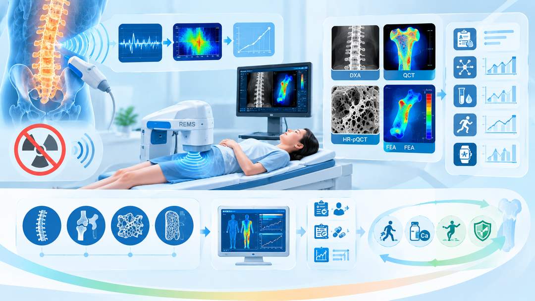
射频回波超声多谱分析(radiofrequency echographic multi-spectrometry,REMS)是一种基于超声射频信号分析的骨密度评估技术,无需使用电离辐射,可用于腰椎和股骨近端等中轴骨骼部位的骨矿物质密度测量。现有研究显示,REMS与双能X线吸收法(DXA)具有较好的诊断一致性,并在骨折风险评估方面展现出潜力。但其临床应用仍需关注设备标准化、不同人群的适用性、骨折预测能力及长期监测性能。REMS有望成为骨质疏松筛查与骨健康管理的重要补充技术,但目前不宜将其简单视为DXA的全面替代方案。
Radiofrequency echographic multi-spectrometry (REMS) is an ultrasound-based technology that analyzes radiofrequency signals to assess bone mineral density without ionizing radiation. It can evaluate axial skeletal sites, including the lumbar spine and proximal femur. Current evidence indicates good diagnostic agreement between REMS and dual-energy X-ray absorptiometry (DXA), with potential advantages in fracture-risk assessment. Nevertheless, device standardization, population-specific applicability, fracture-prediction performance, and long-term monitoring remain important considerations. REMS may become a valuable complementary technology for osteoporosis screening and bone health management, but it should not yet be regarded as a universal replacement for DXA.
一、技术原理:从超声回波到骨密度估算
REMS并非传统的定量超声(QUS)的简单升级,也不是通过超声直接测量骨矿物质的质量。其基本原理是利用超声探头采集骨表面及邻近组织产生的射频回波信号,并通过频谱分析与设备内置的参考数据库进行比较,识别与骨组织结构及骨密度相关的信号特征,进而估算骨矿物质密度和相关骨脆弱性指标。
REMS is not simply an upgraded version of conventional quantitative ultrasound (QUS), nor does it directly measure bone mineral mass using ultrasound. Its underlying principle is to acquire radiofrequency echoes from bone surfaces and surrounding tissues, analyze their spectral characteristics, and compare them with device-specific reference databases. The resulting signal features are used to estimate bone mineral density and related indicators of skeletal fragility.
与DXA使用两种不同能量的X射线估算骨矿物质密度不同,REMS不产生电离辐射,因此适合探索需要减少辐射暴露的骨健康评估场景。临床上,REMS主要应用于腰椎及股骨近端测量。其结果通常包括骨矿物质密度、T值或Z值,以及特定设备提供的骨脆弱性相关参数。不同设备、软件版本及分析算法的输出应依据各自的验证资料进行解释。
Unlike DXA, which estimates bone mineral density using X-rays at two energy levels, REMS does not generate ionizing radiation. This makes it attractive for bone health assessment settings in which radiation exposure should be minimized. Clinically, REMS is primarily used to assess the lumbar spine and proximal femur. Outputs may include BMD, T-scores or Z-scores, and device-specific parameters associated with skeletal fragility. Results should be interpreted according to the validation evidence for the relevant device, software version, and analytical algorithm.
二、诊断准确性与现有临床证据
2025年发表的一篇系统综述纳入了2015年至2024年12月间发表的相关研究。经过筛选,17项研究进入全文评估。总体而言,研究显示REMS与DXA之间具有较好的诊断一致性,并具有良好的操作者内重复性。部分研究还提示,当腰椎DXA受到椎体变形、骨关节炎或血管钙化等因素干扰时,REMS可能提供有价值的补充信息。
A systematic review published in 2025 evaluated studies published between 2015 and December 2024. Seventeen studies were retained for full-text assessment. Overall, the reviewed evidence indicated good diagnostic agreement between REMS and DXA, together with favorable intra-operator repeatability. Some studies also suggested that REMS may provide complementary information when lumbar DXA measurements are affected by vertebral deformities, osteoarthritis, or vascular calcification.
不过,诊断一致性不等于两种技术在所有临床决策中完全等效。DXA具有长期积累的诊断、治疗决策及随访证据,也是目前广泛采用的骨密度评估参考方法。REMS的研究结果还可能受到患者体型、软组织厚度、测量部位、操作技术和设备算法的影响。因此,临床评价不能只关注相关系数或平均差异,还应考察诊断阈值附近的一致性、敏感度、特异度及不同疾病亚组中的表现。
However, diagnostic agreement does not establish complete equivalence between the two technologies for every clinical decision. DXA has a long-established evidence base for diagnosis, treatment decisions, and follow-up, and remains the widely used reference method for BMD assessment. REMS measurements may also be influenced by body habitus, soft-tissue thickness, anatomical site, operator technique, and device algorithms. Clinical evaluation should therefore extend beyond correlation coefficients and mean differences to examine agreement near diagnostic thresholds, sensitivity, specificity, and performance across disease subgroups.
三、骨折风险预测:从骨密度走向骨脆弱性评估
REMS的一个重要探索方向,是将骨密度与骨脆弱性相关信息结合,用于识别潜在高风险人群。部分研究提示,REMS生成的骨脆弱性相关指标与骨折风险有关,联合分析可能优于单独使用骨密度。但这些结果需要结合研究设计、样本量、随访时间和终点定义进行判断,不能据此推断REMS在所有人群中均优于DXA。
An important direction for REMS research is the integration of BMD with bone fragility-related information to identify individuals at elevated risk. Some studies suggest that REMS-derived fragility parameters are associated with fracture risk and that combined assessment may outperform BMD alone. However, these findings must be interpreted in the context of study design, sample size, follow-up duration, and endpoint definitions; they do not establish universal superiority over DXA.
未来应通过前瞻性队列研究,比较REMS、DXA及FRAX等工具对髋部骨折、椎体骨折和主要骨质疏松性骨折的预测效能,并验证其能否改善临床决策。尤其需要明确:REMS增加的预测信息是否独立于年龄、既往脆性骨折、糖皮质激素使用及传统骨密度指标,以及这种增益能否转化为可测量的临床获益。
Future prospective cohort studies should compare REMS with DXA and tools such as FRAX for predicting hip, vertebral, and major osteoporotic fractures, while evaluating whether REMS improves clinical decision-making. In particular, researchers should determine whether the additional predictive information from REMS is independent of age, prior fragility fractures, glucocorticoid exposure, and conventional BMD measurements, and whether any incremental predictive value translates into measurable clinical benefit.
四、临床应用场景与现实局限
无辐射、可在非传统影像环境中开展评估,是REMS具有吸引力的原因之一。它有望应用于骨质疏松初筛、社区骨健康管理、特定人群的重复评估,以及部分不便接受DXA检查者的辅助评估。不过,是否适合某一场景,仍取决于设备可及性、操作人员培训、质量控制和当地监管要求。
The absence of ionizing radiation and the potential for assessment outside conventional imaging environments make REMS attractive. Possible applications include initial osteoporosis screening, community-based bone health programs, repeat assessment in selected populations, and complementary evaluation of individuals who have difficulty accessing DXA. Suitability for each setting nevertheless depends on device availability, operator training, quality assurance, and local regulatory requirements.
临床应用中必须区分“骨密度测量”“骨质疏松诊断”和“骨折风险预测”三个层次。国际临床骨密度测量学会(ISCD)2023年成人官方立场强调,非传统DXA设备应有独立验证依据,不能将任意设备生成的T值直接套用WHO骨质疏松诊断分类。因此,REMS结果应依据相应设备的验证研究、适用人群和当地指南解读;对诊断不明确、存在脆性骨折或需要制定药物治疗方案的患者,应结合临床危险因素,必要时进行DXA或其他适当检查。
Clinical practice must distinguish among BMD measurement, osteoporosis diagnosis, and fracture-risk prediction. The 2023 Adult Official Positions of the International Society for Clinical Densitometry (ISCD) emphasize the need for independent validation of non-DXA technologies and caution against automatically applying WHO diagnostic classifications to T-scores generated by unvalidated devices. REMS results should therefore be interpreted according to device-specific validation, intended populations, and local guidelines. Patients with uncertain diagnoses, fragility fractures, or a need for treatment decisions require comprehensive clinical assessment and, when indicated, DXA or other appropriate investigations.
五、未来研究方向与结论
REMS下一阶段的关键任务,是建立跨设备、跨人群的标准化验证体系,明确不同性别、年龄、体型及疾病状态下的测量性能,并开展长期随访和临床结局研究。还应评估其重复测量的最小显著变化、治疗监测价值、成本效益,以及在基层筛查和骨折二级预防体系中的实际作用。
The next phase of REMS development should establish standardized validation across devices and populations, clarify measurement performance across sex, age, body habitus, and disease states, and conduct long-term follow-up studies with clinical endpoints. Further work should also assess least significant change for serial measurements, treatment-monitoring utility, cost-effectiveness, and practical value in primary-care screening and secondary fracture-prevention programs.
总体而言,REMS是一项具有临床潜力的无电离辐射骨密度评估技术,其价值不仅在于避免辐射暴露,更在于探索骨密度与骨脆弱性联合评估的新路径。现阶段更合理的定位是经过验证后的补充性评估工具,而非DXA的普遍替代方案;其能否改变骨质疏松筛查与治疗实践,最终取决于前瞻性骨折预测证据、标准化质量控制及临床获益研究。
In summary, REMS is a promising radiation-free technology for bone assessment. Its potential value extends beyond avoiding radiation exposure to exploring integrated assessment of BMD and skeletal fragility. At present, its most appropriate role is as a validated complementary assessment tool rather than a universal replacement for DXA. Whether it can transform osteoporosis screening and treatment will ultimately depend on prospective fracture-prediction evidence, standardized quality assurance, and demonstrated clinical benefit.
参考文献
1. Zambito K, Kushchayeva Y, Bush A, et al. Proposed practice parameters for the performance of radiofrequency echographic multispectrometry (REMS) evaluations. Bone & Joint Open. 2025;6(3):291–297.
2. Radiofrequency Echographic Multi Spectrometry—A Novel Tool in the Diagnosis of Osteoporosis and Prediction of Fragility Fractures: A Systematic Review. Journal of Clinical Medicine. 2025.
3. International Society for Clinical Densitometry (ISCD). 2023 ISCD Official Positions—Adult. 2023.
4. Hans D, Šteňová E, Briot K, et al. Radiofrequency echographic multi-spectrometry for the assessment of bone mineral density and fracture risk: clinical evidence and perspectives.