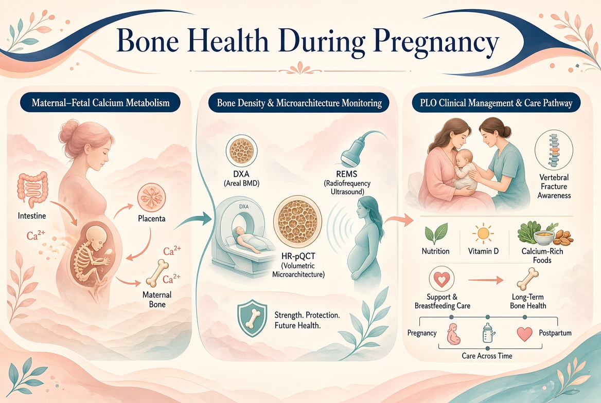
妊娠与哺乳期是女性骨代谢发生显著适应性变化的特殊阶段。胎儿骨骼矿化、母体钙稳态、激素调节及产后骨量恢复相互关联,构成母婴整体健康的重要组成部分。近年来,研究重点逐渐从单纯观察母体骨密度变化,拓展至骨微结构、骨转换机制、营养干预及妊娠与哺乳相关骨质疏松(pregnancy- and lactation-associated osteoporosis,PLO)的早期识别。2026年发布的国际多学科立场声明进一步推动了PLO的规范化评估与个体化管理。本文综述相关研究进展及其临床意义。
Pregnancy and lactation are unique physiological stages characterized by substantial adaptive changes in maternal bone metabolism. Fetal skeletal mineralization, maternal calcium homeostasis, hormonal regulation, and postpartum skeletal recovery are interrelated components of maternal–infant health. Recent research has expanded beyond maternal bone mineral density (BMD) to include bone microarchitecture, remodeling mechanisms, nutritional interventions, and early recognition of pregnancy- and lactation-associated osteoporosis (PLO). An international multidisciplinary position statement published in 2026 further advances structured assessment and individualized management of PLO. This review summarizes these developments and their clinical implications.
一、母胎钙代谢与骨重塑的生理适应
胎儿足月时骨骼约含25–30 g钙,其中大部分在妊娠晚期积累。妊娠期间,母体主要通过提高肠道钙吸收满足胎儿需求;哺乳期则在泌乳素、甲状旁腺激素相关肽(PTHrP)升高及雌激素水平下降等因素作用下,增加骨钙动员。哺乳最初数月腰椎骨密度可出现约5%–10%的生理性下降,通常在断乳后逐步恢复。[1,2]
At term, the fetal skeleton contains approximately 25–30 g of calcium, much of which accumulates during late pregnancy. Maternal adaptation during pregnancy relies primarily on increased intestinal calcium absorption. During lactation, elevated parathyroid hormone-related peptide (PTHrP), prolactin-related endocrine changes, and reduced estrogen levels promote skeletal calcium mobilization. Lumbar spine BMD may decline by approximately 5%–10% during the first months of lactation, with recovery generally occurring after weaning.[1,2]
这种骨量变化不应简单视为病理性骨质疏松。现有证据总体表明,妊娠和哺乳引起的骨量下降多为暂时性变化,不能据此推断所有女性未来骨质疏松或骨折风险增加。临床研究需要同时考察母体营养状况、孕前骨骼基础、泌乳持续时间及断乳后的恢复情况,避免将生理性骨重塑与异常骨脆弱混为一谈。
These changes should not automatically be interpreted as pathological osteoporosis. Overall evidence indicates that pregnancy- and lactation-related bone loss is usually transient; it does not establish that pregnancy or breastfeeding universally increases subsequent osteoporosis or fracture risk. Clinical studies should consider maternal nutritional status, pre-pregnancy skeletal health, lactation duration, and post-weaning recovery to distinguish physiological remodeling from pathological skeletal fragility.
二、骨密度监测与骨微结构评价的新进展
双能X线吸收法(DXA)仍是临床骨密度评价的重要工具,但孕期不宜将其作为常规筛查手段。由于孕妇及胎儿涉及不同的影像安全考量,检查应基于明确的临床指征进行风险—获益评估。对于产后疑似PLO或出现脆性骨折的患者,腰椎和髋部DXA有助于评估骨量状态;对于绝经前女性,应结合年龄、临床表现及Z值解读结果,而不能机械套用绝经后女性的诊断标准。[1,2]
Dual-energy X-ray absorptiometry (DXA) remains an important clinical tool for BMD assessment, but it should not be used for routine screening during pregnancy. Imaging decisions should be based on clear clinical indications and an individualized risk–benefit assessment. In postpartum women with suspected PLO or fragility fractures, lumbar spine and hip DXA can help characterize skeletal status. In premenopausal women, results should be interpreted using age-appropriate Z-scores and the clinical context rather than mechanically applying diagnostic thresholds developed for postmenopausal women.[1,2]
近年来,定量CT、外周定量CT及高分辨率外周定量CT(HR-pQCT)进一步拓展了骨健康研究的评价维度,可用于分析皮质骨、骨小梁结构及体积骨密度。2024年发表的综述总结了妊娠和哺乳期骨量及骨结构变化,指出骨密度并不能完整反映骨强度。无电离辐射的射频超声多频谱技术(REMS)也开始用于妊娠期骨状态研究,但其在孕妇中的验证数据仍有限,尚不能据此认定其已成为常规监测标准。[2,3]
Quantitative CT, peripheral quantitative CT, and high-resolution peripheral quantitative CT (HR-pQCT) have expanded research beyond areal BMD to include cortical and trabecular architecture and volumetric density. A 2024 review highlighted that BMD alone does not fully capture skeletal strength during pregnancy and lactation. Non-ionizing radiofrequency echographic multi-spectrometry (REMS) has also been investigated for assessing bone status during pregnancy, but validation data remain limited, and it cannot yet be considered an established standard for routine monitoring.[2,3]
三、营养干预与母婴整体健康
孕期骨健康管理应纳入整体产前保健,而不是孤立追求骨密度数值。临床评估应关注膳食钙摄入、维生素D状态、蛋白质及总能量摄入、体重变化和相关基础疾病。应优先通过合理膳食满足营养需求,并依据摄入不足、实验室检查和个体风险决定是否补充。对于存在维生素D缺乏、吸收障碍或其他矿物质代谢异常者,应进行针对性纠正;不宜假定额外增加钙剂就能完全阻止哺乳期的生理性骨量下降。[4,5]
Pregnancy-related bone-health management should be integrated into comprehensive antenatal care rather than focused exclusively on BMD values. Assessment should cover dietary calcium, vitamin D status, protein and energy intake, weight changes, and relevant comorbidities. Adequate nutrition should be prioritized, with supplementation guided by dietary insufficiency, laboratory findings, and individual risk. Vitamin D deficiency, malabsorption, and other mineral-metabolism disorders warrant targeted management; additional calcium should not be assumed to prevent all physiological bone loss during lactation.[4,5]
运动干预同样需要兼顾母体安全、妊娠阶段及产科情况。对无禁忌证的孕妇,可根据个体情况安排适度负重活动、肌力训练及平衡练习;对产后女性,则应结合分娩恢复、盆底功能和肌肉骨骼症状逐步恢复活动。未来研究应进一步明确不同运动方案对母体骨微结构、跌倒风险、妊娠结局及婴儿骨骼发育的影响,避免仅以短期骨密度变化作为唯一评价终点。
Exercise interventions should account for maternal safety, gestational stage, and obstetric conditions. For pregnant women without contraindications, individualized weight-bearing activity, strength training, and balance exercises may be appropriate. Postpartum activity should be resumed progressively according to delivery recovery, pelvic-floor function, and musculoskeletal symptoms. Future studies should clarify how different exercise regimens affect maternal bone microarchitecture, fall risk, pregnancy outcomes, and infant skeletal development rather than relying solely on short-term BMD changes.
四、妊娠与哺乳相关骨质疏松:2026年临床管理进展
PLO是一种罕见但可能造成严重后果的疾病,常表现为妊娠晚期或产后早期脆性骨折,以椎体压缩性骨折较为典型。2026年,国际围产医学学会、国际骨质疏松基金会、欧洲钙化组织学会等多个组织联合发布立场声明,建议通过详细病史、体格检查、相关实验室检测及适当影像学检查排查继发性骨质疏松,并根据是否处于妊娠期或产后阶段制定个体化管理方案。[1]
PLO is rare but potentially serious, typically presenting with fragility fractures during late pregnancy or early postpartum, most characteristically vertebral compression fractures. In 2026, a joint position statement from the International Academy of Perinatal Medicine, International Osteoporosis Foundation, European Calcified Tissue Society, and other organizations recommended detailed clinical history, physical examination, laboratory investigations for secondary causes, and appropriate imaging. Management should be individualized according to whether the patient is still pregnant or has entered the postpartum period.[1]
对孕晚期或产后出现持续严重背痛、身高下降或轻微外力后疼痛的患者,应提高对椎体骨折的警惕。处理包括镇痛、纠正营养缺乏、评估哺乳对骨代谢的影响及骨折风险。骨形成促进剂、抗骨吸收药物等应由专科团队个体化权衡;目前缺乏高质量随机对照试验,不能将任何一种药物视为所有患者的标准方案。未来妊娠计划、避孕需求及药物长期安全性均应纳入决策。
Persistent severe back pain, loss of height, or pain following minimal trauma during late pregnancy or postpartum should prompt consideration of vertebral fractures. Management may include analgesia, correction of nutritional deficiencies, and assessment of lactation-related skeletal demands and fracture risk. Osteoanabolic or antiresorptive agents require individualized specialist assessment. High-quality randomized trials remain lacking, so no single drug regimen can be regarded as standard for all patients. Future pregnancy plans, contraception, and long-term medication safety must all inform treatment decisions.
五、总结与未来研究方向
未来孕期骨健康研究需要建立覆盖孕前、妊娠、哺乳及断乳后阶段的纵向队列,联合分析骨密度、骨微结构、骨转换标志物、营养和母婴结局。尤其需要验证非电离辐射骨评价工具的准确性、明确高危人群的合理监测频率,并建立PLO多中心登记和长期随访体系。临床目标不是消除所有生理性骨量变化,而是在保障胎儿发育和母乳喂养的同时,及时识别病理性骨脆弱并降低严重骨折风险。
Future research should establish longitudinal cohorts spanning preconception, pregnancy, lactation, and post-weaning, integrating BMD, bone microarchitecture, turnover markers, nutrition, and maternal–infant outcomes. Priorities include validating non-ionizing bone-assessment technologies, defining appropriate monitoring intervals for high-risk groups, and developing multicenter registries with long-term follow-up for PLO. The clinical objective is not to eliminate all physiological bone loss, but to support fetal development and breastfeeding while identifying pathological skeletal fragility early and reducing the risk of serious fractures.
参考文献
1. Hadji P, Athanasiadis A, Brandi ML, et al. Pregnancy- and lactation-associated osteoporosis: A position statement of the IAPM, IOF, ECTS, ESCEO, IMS, and EMAS. International Journal of Gynecology & Obstetrics. 2026.
2. Cohen A. Bone Metabolism, Bone Mass, and Bone Structure During Pregnancy and Lactation: Normal Physiology and Pregnancy and Lactation-Associated Osteoporosis. Endocrinology and Metabolism Clinics of North America. 2024;53(3):453–470.
3. Brona A, Stachowska B, Bolanowski M, Halupczok-Żyła J. The influence of hormonal changes during pregnancy on bone metabolism—a mini review. Frontiers in Endocrinology. 2026;17.
4. Kovacs CS. Pregnancy and Lactation Associated Bone Fragility. Journal of the Endocrine Society. 2025;9(9):bvaf126.
5. National Institute of Arthritis and Musculoskeletal and Skin Diseases. Pregnancy, Breastfeeding, and Bone Health.
注:本文为叙述性综述,文献更新截至2026年10月。孕期骨密度检查、营养补充及PLO治疗均应根据具体临床指征和个体风险决定。