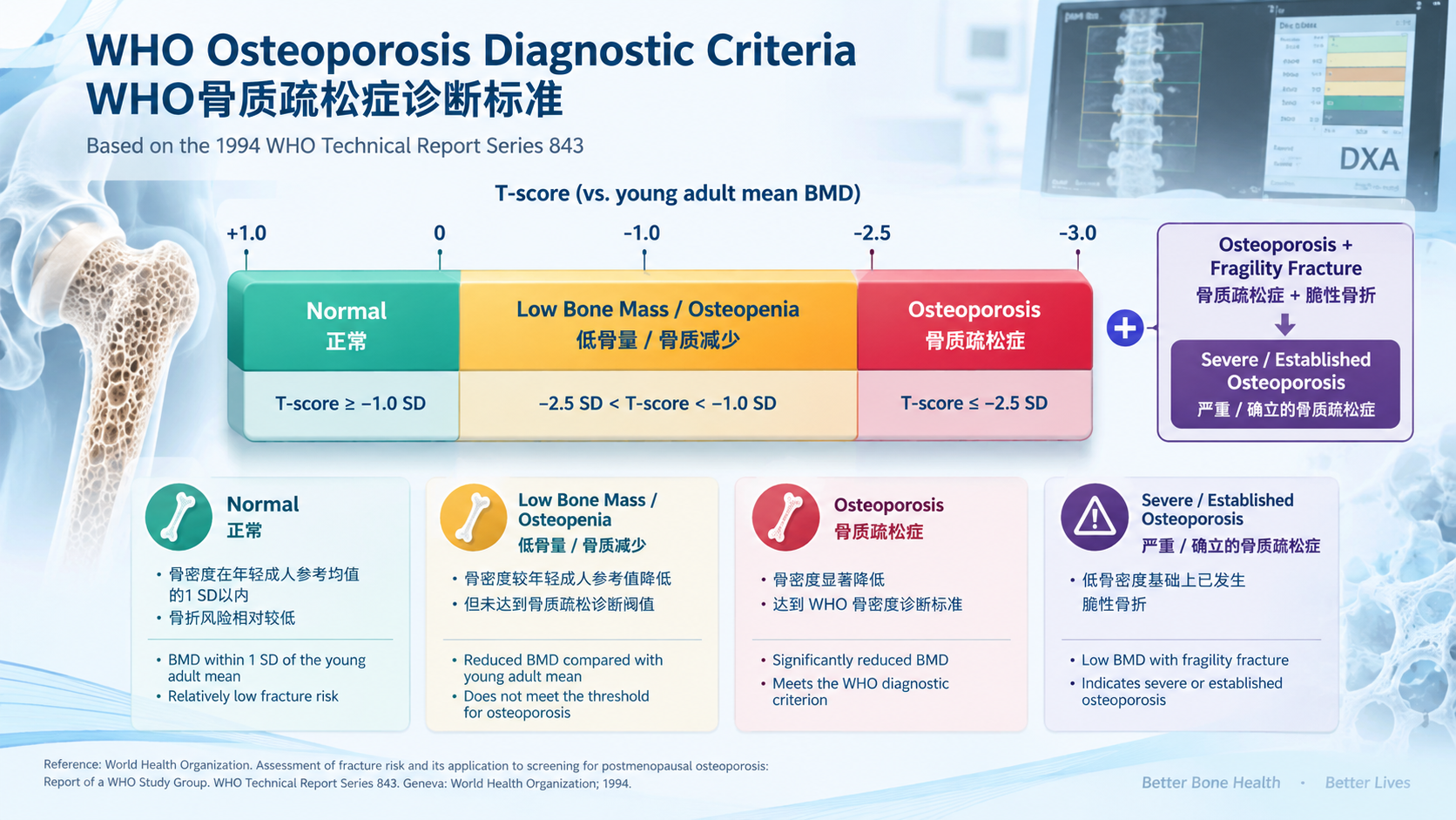
1. 标准概述|Overview
世界卫生组织(WHO)骨质疏松症诊断标准的核心是基于骨密度(bone mineral density,BMD)的T-score。1994年WHO研究组提出以年轻成年女性峰值骨量为参照,将骨密度降低程度进行操作性分类,其中T-score ≤−2.5 SD被定义为骨质疏松症。该标准最初主要针对绝经后女性建立,随后被国际骨密度测量与骨质疏松诊疗实践广泛采用,并成为全球最具影响力的骨质疏松骨密度分类体系之一。
The World Health Organization (WHO) operational definition of osteoporosis is primarily based on bone mineral density (BMD) expressed as a T-score. In 1994, the WHO Study Group proposed a classification based on the difference between an individual's BMD and that of a healthy young adult reference population. A T-score of ≤−2.5 standard deviations (SD) was defined as osteoporosis. Although the original framework was developed mainly for postmenopausal women, it subsequently became the internationally recognized densitometric classification used widely in clinical practice.
2. WHO骨密度分类|WHO BMD Classification
按照经典WHO骨密度分类,T-score ≥−1.0 SD属于正常骨密度;T-score介于−1.0和−2.5 SD之间属于低骨量(low bone mass),传统上称为“骨量减少”或“骨质减少(osteopenia)”;T-score ≤−2.5 SD则达到骨质疏松症的骨密度诊断标准。在早期WHO分类中,如果患者同时存在骨质疏松范围的BMD和脆性骨折,则可描述为“严重骨质疏松症(severe or established osteoporosis)”。
Under the classic WHO densitometric classification, a T-score of ≥−1.0 SD is considered normal. A T-score between −1.0 and −2.5 SD indicates low bone mass, traditionally termed osteopenia, whereas a T-score of ≤−2.5 SD meets the densitometric criterion for osteoporosis. The original WHO framework also described severe or established osteoporosis when osteoporosis-range BMD was accompanied by a fragility fracture.
3. T-score的定义|Definition of the T-score
T-score表示受检者BMD与年轻成年参考人群平均BMD之间的标准差距离,其计算形式为:T-score =(患者BMD-年轻成人平均BMD)/年轻成人BMD标准差。需要注意,T-score并不是“骨密度百分比”,而是一个标准化统计指标。因此,T-score下降1个单位并不意味着BMD下降10%或其他固定比例。
The T-score represents the number of standard deviations by which an individual's BMD differs from the mean BMD of a young adult reference population. It is calculated as: T-score = (patient BMD − young-adult mean BMD) / young-adult BMD SD. The T-score is therefore a standardized statistical measure rather than a percentage of bone density, and a one-unit decrease in T-score does not correspond to a fixed percentage reduction in BMD.
4. 推荐的DXA测量部位|Recommended DXA Measurement Sites
现代临床实践中,中央双能X线吸收法(dual-energy X-ray absorptiometry,DXA)是骨质疏松骨密度诊断的标准方法。根据2023年国际临床骨密度学会(ISCD)成人官方立场,绝经后女性和50岁及以上男性可以根据腰椎、全髋或股骨颈的T-score进行WHO骨密度分类;在特定情况下,非优势前臂33%桡骨(one-third/33% radius)也可用于诊断。髋部诊断通常采用股骨颈或全髋中较低的有效T-score。
Central dual-energy X-ray absorptiometry (DXA) is the standard technique for densitometric assessment. According to the 2023 International Society for Clinical Densitometry (ISCD) Official Positions, the WHO densitometric classification can be applied to the lumbar spine, total hip, or femoral neck in postmenopausal women and men aged 50 years or older. The 33% radius may be used for diagnosis in selected circumstances. For the hip, the lower valid T-score from the femoral neck or total hip is generally used for diagnostic classification.
5. WHO标准与DXA的关系|WHO Criteria and DXA
需要强调,WHO的T-score诊断分类并不是可以直接套用到所有骨密度检测技术上的统一阈值。ISCD明确指出,WHO诊断分类主要适用于经过验证的DXA测量部位;来自定量CT(QCT)、外周QCT、定量超声(QUS)或其他设备的T-score不能简单视为与中央DXA T-score等价。不同设备之间也不能直接比较BMD数值。
Importantly, the WHO T-score threshold should not be indiscriminately applied to every bone assessment technology. The ISCD specifies that the WHO diagnostic classification is applicable primarily to validated DXA measurement sites. T-scores derived from quantitative CT (QCT), peripheral QCT, quantitative ultrasound (QUS), or other technologies are not automatically equivalent to central DXA T-scores, and BMD values obtained from different devices should not be directly compared.
6. 不同年龄人群的T-score与Z-score|T-scores versus Z-scores
WHO骨密度分类主要适用于绝经后女性以及50岁及以上男性。对于绝经前女性、50岁以下男性以及儿童和青少年,临床报告通常应优先采用Z-score,而不是T-score。2023年ISCD建议,Z-score ≤−2.0可描述为“低于同年龄预期范围(below the expected range for age)”,但不能仅凭这一数值直接套用WHO绝经后骨质疏松诊断标准。
The WHO densitometric classification is primarily intended for postmenopausal women and men aged 50 years or older. For premenopausal women, men younger than 50 years, children, and adolescents, Z-scores are generally preferred. The 2023 ISCD Official Positions define a Z-score of ≤−2.0 as “below the expected range for age.” A low Z-score alone should not be interpreted as equivalent to a WHO diagnosis of osteoporosis in postmenopausal adults.
7. T-score参考数据库|Reference Database for T-scores
T-score的结果高度依赖参考数据库,因此临床报告不能只关注一个数值而忽略参考标准。ISCD目前建议,T-score采用统一的非种族/民族调整女性参考数据库;对于股骨颈和全髋,制造商继续采用NHANES III数据作为参考标准。腰椎T-score则通常采用设备制造商相应的参考数据库。
The calculated T-score depends substantially on the reference database. The ISCD currently recommends a uniform, non-race/ethnicity-adjusted female normative database for T-score calculation. For the femoral neck and total hip, manufacturers should continue to use the NHANES III reference data, while lumbar spine T-scores generally use the manufacturer's appropriate reference database.
8. 骨折并不等同于T-score≤−2.5|Fracture Is Not Equivalent to a T-score ≤−2.5
临床诊断不能仅依赖T-score。大量研究表明,脆性骨折可以发生在T-score高于−2.5 SD的人群中,因此“T-score未达到−2.5”并不能排除骨骼脆弱性。2024年国际DXA最佳实践工作组指出,大多数脆性骨折患者的T-score实际上高于−2.5;成人发生脆性骨折本身就是未来骨折风险的重要预测因素,应进一步评估潜在骨质疏松及其他危险因素。
Clinical assessment should not rely exclusively on the T-score. Fragility fractures frequently occur in individuals whose BMD T-score is above −2.5, meaning that a T-score above the osteoporosis threshold does not exclude skeletal fragility. A 2024 international working group emphasized that most patients with fragility fractures have T-scores higher than −2.5 and that an adult fragility fracture should prompt further evaluation for osteoporosis and other fracture risk factors.
9. FRAX与骨质疏松诊断|FRAX and Osteoporosis Diagnosis
WHO T-score标准主要解决“骨密度是否达到骨质疏松范围”的分类问题,而FRAX等工具则用于估计未来一定时期内发生骨折的概率,两者并非完全相同的概念。临床决策还应结合年龄、既往脆性骨折、糖皮质激素使用、父母髋部骨折史、吸烟、饮酒、类风湿关节炎及其他继发性骨质疏松因素等进行综合判断。
The WHO T-score criterion primarily provides a densitometric classification, whereas tools such as FRAX estimate an individual's probability of sustaining a fracture over a defined period. These concepts should not be regarded as interchangeable. Clinical assessment should integrate age, previous fragility fracture, glucocorticoid exposure, parental hip fracture, smoking, alcohol consumption, rheumatoid arthritis, and other causes of secondary osteoporosis.
10. 椎体骨折与隐匿性骨折评估|Vertebral Fracture Assessment
椎体骨折是骨质疏松临床评估中容易被低估的部分。许多低能量椎体骨折没有明显急性疼痛或典型外伤史,因此单纯依赖患者症状可能造成漏诊。DXA结合椎体骨折评估(vertebral fracture assessment,VFA)或必要时进行脊柱X线检查,可以帮助发现影像学椎体骨折。近年来国际DXA最佳实践进一步强调了VFA在骨质疏松评估中的价值。
Vertebral fractures are an important but frequently underrecognized component of osteoporosis assessment. Many low-trauma vertebral fractures are clinically silent and may not present with acute pain or a clear history of trauma. DXA-based vertebral fracture assessment (VFA), or conventional spinal radiography when appropriate, can improve detection. Recent international DXA best-practice recommendations have further emphasized the role of VFA in comprehensive fracture-risk assessment.
11. WHO标准的局限性与现代发展|Limitations and Contemporary Developments
WHO的−2.5 SD阈值具有重要的历史和临床价值,但并不意味着所有人群具有完全相同的骨折风险。近年来研究开始重新讨论不同种族、地区及人群应用统一T-score阈值的适用性。例如,2023年的研究指出,将1994年WHO标准直接应用于部分东亚老年人群,可能导致骨质疏松患病率与实际脆性骨折风险之间出现偏差。不过,目前国际临床实践仍以WHO/ISCD体系作为最主要的骨密度分类框架,各地区应同时遵循本国或地区指南。
The −2.5 SD threshold remains highly influential, but it does not imply that identical T-scores correspond to exactly the same fracture risk across all populations. Recent research has questioned whether the original threshold should be applied uniformly across different ethnic and geographic populations. For example, a 2023 analysis suggested that direct application of the 1994 WHO threshold to some older East Asian populations may overestimate osteoporosis prevalence relative to observed fragility fracture risk. Nevertheless, the WHO/ISCD framework remains the principal international system for densitometric classification, while local clinical guidelines should also be considered.
12. DXA、QCT及新型评估方法|DXA, QCT and Emerging Assessment Methods
DXA仍是骨质疏松骨密度诊断的核心技术,但QCT等三维定量技术能够测量体积骨密度,并可能识别更多低骨密度患者。2025年一项系统综述和Meta分析纳入19项研究、3939例受试者,发现QCT在同一人群中识别出的骨质疏松患者明显多于DXA。由于两种技术测量原理、参考标准和诊断阈值不同,这一结果并不意味着可以直接用QCT替代WHO DXA T-score标准。
DXA remains the central technology for densitometric diagnosis, while three-dimensional techniques such as QCT can provide volumetric BMD and may identify more individuals with low bone density. A 2025 systematic review and meta-analysis including 19 studies and 3,939 participants found that QCT identified substantially more patients as having osteoporosis than DXA. However, this difference reflects the distinct measurement principles and thresholds of the two techniques and does not mean that QCT measurements can simply replace the WHO DXA T-score classification.
13. 临床资料库检索要点|Key Database Retrieval Terms
对于医学专业资料库,建议将本主题与以下关键词交叉检索:WHO osteoporosis criteria、T-score、Z-score、DXA、BMD、femoral neck、total hip、lumbar spine、33% radius、postmenopausal osteoporosis、fragility fracture、vertebral fracture、FRAX、QCT、VFA、ISCD Official Positions。其中,1994年WHO报告和Kanis等人的经典论文适合作为标准历史来源;2023年ISCD官方立场可作为当前DXA报告和T-score解释的重要实践依据;2024—2025年的DXA、脆性骨折及诊断研究则可用于补充现代临床实践背景。
For professional database retrieval, useful search terms include WHO osteoporosis criteria, T-score, Z-score, DXA, BMD, femoral neck, total hip, lumbar spine, 33% radius, postmenopausal osteoporosis, fragility fracture, vertebral fracture, FRAX, QCT, VFA, and ISCD Official Positions. The original 1994 WHO report and the classic paper by Kanis et al. provide historical foundations; the 2023 ISCD Official Positions provide contemporary guidance for DXA interpretation and T-score reporting; and recent studies from 2024–2025 provide updated perspectives on fragility fractures, DXA practice, and alternative diagnostic technologies.
14. 核心分类速查|Quick Reference
|
T-score |
WHO传统分类 |
Clinical category |
|
≥ −1.0 |
正常 |
Normal |
|
−1.0 ~ > −2.5 |
低骨量/骨质减少 |
Low bone mass / Osteopenia |
|
≤ −2.5 |
骨质疏松症 |
Osteoporosis |
|
≤ −2.5 + 脆性骨折 |
严重/确立的骨质疏松症(经典WHO术语) |
Severe / established osteoporosis |
需要注意,上述表格是骨密度分类标准,并不等同于完整的临床骨折风险分层或药物治疗适应证。实际诊疗应结合脆性骨折史、临床危险因素、继发性骨质疏松评估以及FRAX等工具,并依据所在国家或地区的现行指南制定治疗决策。2023年ISCD也明确区分了“诊断分类”和“骨折风险评估”两个概念。
The table above represents a densitometric classification, not a complete clinical fracture-risk or treatment classification. Clinical decision-making should incorporate previous fragility fractures, clinical risk factors, evaluation for secondary osteoporosis, and validated fracture-risk tools such as FRAX, together with applicable national or regional guidelines. The 2023 ISCD Official Positions explicitly distinguish diagnostic classification from fracture-risk assessment.
参考文献|References
1. World Health Organization. Assessment of fracture risk and its application to screening for postmenopausal osteoporosis: Report of a WHO Study Group. WHO Technical Report Series 843. Geneva: World Health Organization; 1994.
2. Kanis JA, Melton LJ 3rd, Christiansen C, Johnston CC, Khaltaev N. The diagnosis of osteoporosis. Journal of Bone and Mineral Research. 1994;9(8):1137-1141. doi:10.1002/jbmr.5650090802.
3. International Society for Clinical Densitometry (ISCD). Official Positions 2023: Adult. ISCD; 2023.
4. Khan AA, Slart RHJA, Ali DS, et al. Osteoporotic Fractures: Diagnosis, Evaluation, and Significance From the International Working Group on DXA Best Practices. Mayo Clinic Proceedings. 2024;99(7):1127-1141. doi:10.1016/j.mayocp.2024.01.011.
5. Yang J, Zeng Y, Yu W. Criteria for osteoporosis diagnosis: a systematic review and meta-analysis of osteoporosis diagnostic studies with DXA and QCT. EClinicalMedicine. 2025;83:103244. doi:10.1016/j.eclinm.2025.103244.
6. Brandt IAG, et al. Diagnosing Osteoporosis in Diabetes—A Systematic Review on BMD and Fractures. Current Osteoporosis Reports. 2024;22(2):223-244. doi:10.1007/s11914-024-00867-1.
7. Revision of the 1994 World Health Organization T-score definition of osteoporosis for use in older East Asian women and men to reconcile it with their lifetime risk of fragility fracture. Osteoporosis International. 2023.
8. Updated practice guideline for dual-energy X-ray absorptiometry (DXA). 2024.