引言|Introduction
双能X线吸收法(dual-energy X-ray absorptiometry,DXA)仍是临床评价骨矿物质密度(bone mineral density,BMD)、诊断骨质疏松以及监测治疗反应的核心工具。但DXA报告并不是简单地“看一个T值”:真正有临床意义的解读,需要同时关注测量部位、BMD绝对值、T-score或Z-score、技术质量、椎体可评估性、骨折风险以及随访时的最小显著变化(least significant change,LSC)。2023年ISCD官方立场进一步强调,DXA的诊断分类与骨折风险评估并不是完全相同的概念。
Dual-energy X-ray absorptiometry (DXA) remains the central clinical tool for assessing bone mineral density (BMD), diagnosing osteoporosis, and monitoring treatment response. However, interpretation should never be reduced to a single T-score. A clinically meaningful DXA interpretation requires assessment of the skeletal site, absolute BMD, T- or Z-score, technical quality, vertebral evaluability, fracture risk, and least significant change (LSC) during follow-up. The 2023 ISCD Official Positions emphasize that densitometric diagnosis and fracture-risk assessment are related but distinct clinical concepts.
1. 先看测量部位,而不是先看T-score|Start With the Skeletal Site, Not the T-score
标准中央DXA通常包括腰椎和髋部。腰椎一般采用后前位(PA)L1–L4;髋部主要分析股骨颈(femoral neck)和全髋(total hip)。对于绝经后女性以及≥50岁的男性,WHO骨密度诊断标准可应用于腰椎、股骨颈或全髋;如果双侧髋均进行了检测,诊断分类应采用有效ROI中最低的股骨颈或全髋T-score,而不是两侧平均值。前臂并非所有患者的常规诊断部位,在髋部或脊柱无法可靠测量、甲状旁腺功能亢进或患者体重超过DXA检查床限制时尤其有价值,此时通常采用非优势侧33%桡骨。
A standard central DXA examination generally includes the lumbar spine and hip. The lumbar spine is usually assessed at PA L1–L4, while the hip is evaluated primarily using the femoral neck and total hip regions of interest. In postmenopausal women and men aged 50 years or older, WHO densitometric criteria may be applied to the lumbar spine, femoral neck, or total hip. When both hips are measured, the lowest valid femoral-neck or total-hip T-score should be used for diagnostic classification rather than averaging the two sides. The 33% radius is particularly useful when the hip or spine cannot be interpreted, in hyperparathyroidism, or when the patient exceeds the DXA table weight limit.
2. BMD、T-score与Z-score必须分开理解|Distinguish BMD, T-score, and Z-score
BMD以g/cm²表示,是DXA最直接的测量结果。T-score则表示患者BMD与年轻成年参考人群峰值骨量之间相差多少个标准差(SD);Z-score则是与年龄匹配人群相比的标准差距离。对于绝经后女性和≥50岁男性,T-score是主要诊断指标;T-score ≥−1.0通常归为正常,−1.0至−2.5之间为低骨量,≤−2.5达到骨质疏松的骨密度标准。对于绝经前女性和50岁以下男性,则优先使用Z-score;Z-score ≤−2.0通常表述为“低于同龄人预期范围”,而不是直接套用绝经后人群的T-score诊断标准。
BMD, expressed in g/cm², is the primary quantitative measurement produced by
DXA. The T-score represents the number of standard deviations by which a
patient's BMD differs from the young-adult reference population, whereas the
Z-score compares BMD with an age-matched reference population. T-scores are
preferred for postmenopausal women and men aged 50 years or older. A T-score of
−1.0 or above is generally considered normal, between −1.0 and −2.5 indicates
low bone mass, and ≤−2.5 meets the densitometric criterion for osteoporosis. In
premenopausal women and men younger than 50 years, Z-scores are preferred; a
Z-score ≤−2.0 is described as “below the expected range for age” rather than
being used alone to diagnose osteoporosis.
3. “最低T-score”很重要,但不能脱离临床背景|The Lowest T-score Matters—but Not in Isolation
临床实践中常见的错误是看到某一个部位T-score最低,就直接将其等同于患者整体骨折风险。例如腰椎T-score为−2.7而髋部为−1.8,患者满足骨密度学骨质疏松标准,但其实际骨折风险还需要结合年龄、既往脆性骨折、糖皮质激素暴露、吸烟、饮酒、继发性骨质疏松因素以及其他危险因素综合判断。因此,“T-score≤−2.5”是诊断框架的重要组成部分,却并不等于“已经完整完成骨折风险评估”。
A common interpretive error is to equate the lowest T-score with the patient's
overall fracture risk. For example, a lumbar-spine T-score of −2.7 with a hip
T-score of −1.8 fulfills the densitometric criterion for osteoporosis, but
fracture risk still depends on age, previous fragility fractures,
glucocorticoid exposure, smoking, alcohol use, secondary causes of
osteoporosis, and other clinical risk factors. Thus, a T-score ≤−2.5 is an
important diagnostic threshold, but it does not constitute a complete fracture-risk
assessment.
4. 仔细检查腰椎:退行性改变可能“虚高”BMD|Inspect the Lumbar Spine Carefully: Degeneration Can Falsely Elevate
BMD
腰椎BMD特别容易受到非骨质疏松因素干扰。椎体骨赘、终板硬化、椎间隙改变、椎体压缩、骨折、椎体成形术以及金属内固定等均可能造成局部BMD升高。如果某一椎体明显异常,或者其T-score与相邻椎体相差超过1.0,应考虑排除该椎体后重新计算结果。值得注意的是,不能仅凭单个椎体诊断或监测骨质疏松;2023年ISCD进一步强调,当可用椎体少于4个时,应结合本机构精密度数据重新确定相应LSC。
Lumbar-spine BMD is particularly vulnerable to artifacts unrelated to bone
strength. Osteophytes, endplate sclerosis, degenerative disc disease, vertebral
compression, fractures, vertebroplasty, and spinal hardware can artificially
increase measured BMD. A vertebra may be excluded when it is clearly abnormal
or when its T-score differs by more than 1.0 from adjacent vertebrae.
Importantly, a single vertebral body should not be used for osteoporosis
diagnosis or monitoring. When fewer than four vertebrae remain evaluable,
precision and the corresponding LSC should be reassessed according to the
facility's own precision data.
5. DXA不能替代骨折史:VFA尤其值得关注|DXA Does Not Replace Fracture
History: Pay Attention to VFA
椎体骨折是未来骨折风险的重要预测因素,但临床上相当一部分椎体骨折并没有典型急性症状。因此,对于符合条件的患者,应关注报告中是否进行了椎体骨折评估(vertebral fracture assessment,VFA)。ISCD建议,在T-score<−1.0并合并高龄、明显身高下降、既往疑似椎体骨折或长期糖皮质激素治疗等情况下考虑VFA。VFA发现的椎体形态异常不能简单依赖自动形态测量,需要结合影像学表现和临床背景判断。
Vertebral fractures are powerful predictors of future fracture risk, yet many
vertebral fractures remain clinically silent. Therefore, clinicians should
determine whether vertebral fracture assessment (VFA) was indicated and
performed. ISCD recommends considering VFA in patients with a T-score below
−1.0 who also have relevant clinical risk factors, such as advanced age,
substantial historical height loss, an undocumented vertebral fracture, or
prolonged glucocorticoid exposure. Vertebral deformities identified by VFA
should not be interpreted solely by automated morphometry; visual assessment
and clinical context remain essential.
6. 纵向随访最关键的是LSC,而不是“百分比变化”|During Follow-up, LSC Matters More Than Percentage Change Alone
治疗随访时,报告中常见“BMD较前增加3%”或“下降2%”,但这种变化是否具有统计学意义,不能仅凭百分比判断。必须知道检测中心自身的精密度误差以及LSC。如果变化未超过LSC,就不应简单描述为“骨量下降”或“治疗无效”。此外,随访最好使用同一台DXA设备;不同设备之间如果没有完成适当的交叉校准,不应直接比较BMD变化。
During longitudinal monitoring, reports often state that BMD has increased by
3% or decreased by 2%. Such a change cannot be interpreted solely from the
percentage difference. The facility-specific precision error and least
significant change (LSC) must be considered. A change that does not exceed the
LSC should not automatically be labeled as significant bone loss or treatment
failure. Follow-up measurements should preferably be performed on the same DXA
system; comparisons between non-cross-calibrated instruments should not be
interpreted as quantitative change.
7. 不要忽略FRAX和TBS:从“骨密度”走向“骨折风险”|Do Not Ignore FRAX and TBS: Moving From BMD to Fracture Risk
DXA回答的是“骨密度是多少”,而临床决策更关心“患者未来发生骨折的概率是多少”。因此,在适当人群中,应将BMD与FRAX等临床风险评估工具结合。对于部分接近治疗决策阈值的患者,TBS(trabecular bone score)可以作为骨微结构相关的信息,并用于调整FRAX风险。但TBS不能脱离BMD、临床危险因素和既往骨折史单独决定治疗。
DXA primarily answers the question, “How low is the patient's BMD?” whereas
clinical management is ultimately concerned with “What is the patient's
probability of future fracture?” Therefore, BMD should be integrated with
validated clinical risk-assessment tools such as FRAX when appropriate. In
selected patients close to a treatment threshold, trabecular bone score (TBS)
may provide additional information related to trabecular texture and can be
incorporated into FRAX adjustment. However, TBS should not be used as a
stand-alone determinant of pharmacological treatment.
8. 一个实用的DXA报告阅读顺序|A Practical Sequence for Reading a DXA Report
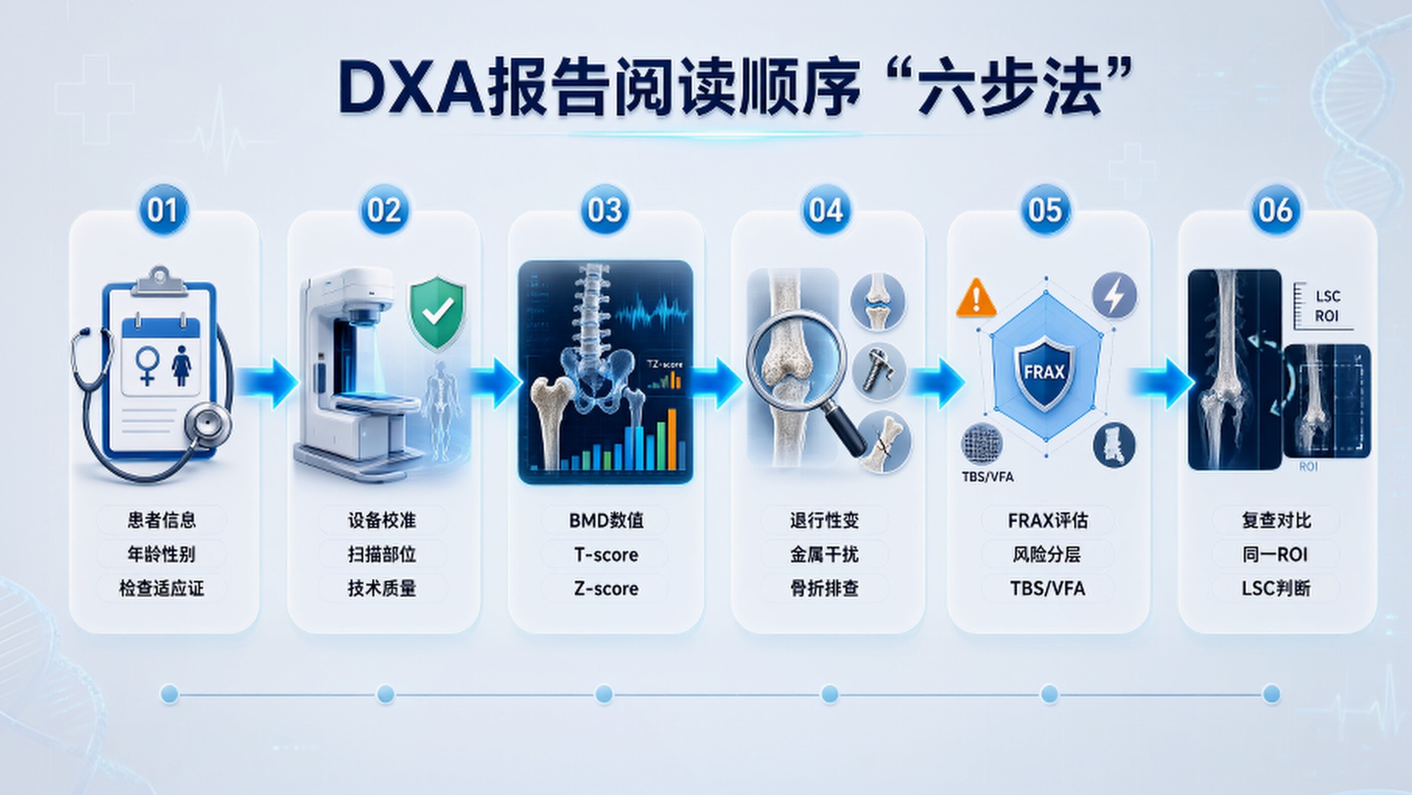
对于临床医生,可以采用“六步法”:①确认患者年龄、性别及检查适应证;②检查DXA设备、扫描部位和技术质量;③查看腰椎、股骨颈和全髋的BMD及T/Z-score;④判断是否存在退行性改变、金属、骨折等影响因素;⑤结合既往脆性骨折、危险因素、FRAX和必要时的TBS/VFA进行整体风险分层;⑥如果是复查,则比较同一设备、同一ROI下的BMD变化,并判断是否超过LSC。只有完成这六个步骤,DXA报告才真正从“一张数字表”转化为临床决策工具。
A practical six-step approach is useful: (1) confirm age, sex, and the clinical
indication; (2) review the DXA system, skeletal sites, and technical quality;
(3) examine BMD and T/Z-scores at the spine, femoral neck, and total hip; (4)
identify degenerative changes, hardware, fractures, or other artifacts; (5)
integrate prior fragility fractures, clinical risk factors, FRAX, and, when
appropriate, TBS and VFA; and (6) for follow-up studies, compare BMD using the
same system and ROI and determine whether the change exceeds the
facility-specific LSC. This approach transforms DXA from a numerical report
into a clinically meaningful decision-support tool.
结语|Conclusion
高质量DXA解读的核心不是寻找“最低的一个数字”,而是建立从测量质量→BMD→T/Z-score→影像学异常→骨折史→FRAX/TBS→纵向变化→临床决策的完整逻辑链。对于医学专业人员而言,理解DXA报告中的每一个数字如何产生、哪些数字可以比较、哪些变化具有统计学意义,比单纯记住“−2.5就是骨质疏松”更加重要。
The essence of high-quality DXA interpretation is not to identify the single
lowest number, but to construct a complete clinical reasoning pathway: measurement
quality → BMD → T/Z-score → imaging abnormalities → fracture history → FRAX/TBS
→ longitudinal change → clinical decision-making. For healthcare
professionals, understanding how each DXA value is generated, which
measurements are comparable, and which changes are statistically significant is
far more valuable than simply memorizing that “−2.5 means osteoporosis.”
参考文献|References
- International Society for Clinical Densitometry (ISCD). 2023 Official Positions—Adult. ISCD; 2023.
- Riemer HJA Slart, Punda M, Ali DS, et al. Updated practice guideline for dual-energy X-ray absorptiometry (DXA). European Journal of Nuclear Medicine and Molecular Imaging. 2025;52(2):539–563. doi:10.1007/s00259-024-06912-6.
- Rosen H, Lau A, Szalat A, et al. Reporting Fewer Than Four Vertebrae: 2023 Official Positions of the International Society for Clinical Densitometry. Journal of Clinical Densitometry. 2024;27(1):101436. doi:10.1016/j.jocd.2023.101436.
- Kanis JA, Johnell O, Oden A, et al. FRAX and the assessment of fracture probability in men and women from the UK. Osteoporosis International. 2008;19:385–397.
- Genant HK, Wu CY, van Kuijk C, Nevitt MC. Vertebral fracture assessment using a semiquantitative technique. Journal of Bone and Mineral Research. 1993;8:1137–1148.
- Hans D, Goertzen AL, Krieg MA, Leslie WD. Bone microarchitecture and the role of trabecular bone score (TBS) in fracture risk assessment. Osteoporosis International. 2011;22:163–172.
- Cummings SR, Bates D, Black DM. Clinical use of bone densitometry: scientific review. JAMA. 2002;288:1889–1897.