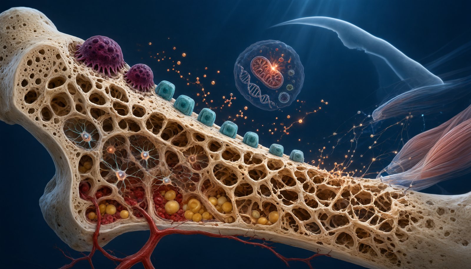
衰老并不只是让骨骼“变轻”,而是会从分子、细胞、组织和器官多个层面改变骨骼的结构与功能。成年以后,骨组织仍然持续进行骨重塑(bone remodeling):破骨细胞清除旧骨,成骨细胞形成新骨,骨细胞则感受机械刺激并协调这一过程。随着年龄增长,骨吸收与骨形成之间的平衡逐渐发生改变,骨量下降、骨小梁变薄和断裂、皮质骨孔隙增加以及骨组织力学性能下降,最终使骨骼更容易发生脆性骨折。2026年的一篇最新综述进一步强调,骨衰老并不是单纯的“骨密度下降”,而是一个涉及分子、细胞、微环境和全身系统的多尺度退化过程。
Aging does not simply make bones “lighter.” It progressively changes skeletal structure and function at molecular, cellular, tissue, and systemic levels. Throughout adulthood, bone continuously undergoes bone remodeling, in which osteoclasts remove old bone, osteoblasts form new bone, and osteocytes sense mechanical signals and coordinate the process. With advancing age, the balance between resorption and formation gradually shifts. Trabeculae become thinner and more disconnected, cortical bone becomes increasingly porous, and the mechanical properties of bone deteriorate. These changes ultimately increase susceptibility to fragility fractures. A major 2026 review emphasized that bone aging is not merely a decline in bone mineral density, but a multiscale degenerative process involving molecular, cellular, microenvironmental, and systemic changes.
1. 峰值骨量决定了衰老的“起点”
人的骨量并不是成年以后才开始建立。儿童和青少年时期,骨骼处于快速生长阶段,骨形成明显超过骨吸收;进入成年早期后,骨量达到所谓的峰值骨量(peak bone mass)。此后,随着年龄增加,骨形成能力逐渐下降,而骨吸收可能逐渐占据优势。因此,一个人在年轻时期建立的峰值骨量,可以理解为衰老过程中骨骼的“储备”。遗传因素、营养、身体活动、青春期发育以及激素水平都会影响这一阶段的骨骼积累。
Bone mass is not established only in adulthood. During childhood and adolescence, the skeleton undergoes rapid growth, with bone formation substantially exceeding resorption. In early adulthood, bone mass reaches its peak bone mass, after which age-related changes gradually become more apparent. As aging progresses, bone-forming capacity declines and resorption may increasingly dominate. Peak bone mass can therefore be viewed as an important skeletal “reserve” for later life. Genetics, nutrition, physical activity, pubertal development, and hormonal status all influence how much bone is accumulated during this critical period.
2. 衰老后,骨重塑逐渐失去平衡
年轻骨骼中的骨重塑通常具有较好的耦联关系:一个骨重塑单位完成骨吸收后,随后能够启动相对协调的骨形成。然而,随着年龄增长,成骨细胞的数量和功能可能下降,骨髓间充质干/基质细胞(BMSCs)的成骨潜能降低,同时破骨细胞及其前体受到炎症和微环境改变的影响。最终,部分骨重塑单位可能表现为“吸收多、形成少”,使每一次重塑周期都产生轻微的骨量亏损。
In younger bone, remodeling is generally well coupled: after a remodeling unit removes old bone, subsequent bone formation largely replaces the resorbed tissue. With aging, however, osteoblast number and function decline, while bone marrow mesenchymal stem/stromal cells (BMSCs) lose part of their osteogenic potential. At the same time, osteoclast precursors can be influenced by inflammatory and microenvironmental changes. Some remodeling units therefore become increasingly imbalanced, with more bone removed than replaced, producing a small negative balance after each remodeling cycle.
3. 骨小梁会发生什么变化?
松质骨中的**骨小梁(trabeculae)**是衰老过程中非常敏感的结构。年轻骨骼中的骨小梁相互连接,形成类似三维网状结构,可以有效承受和传递机械负荷。随着年龄增加,部分骨小梁会变薄、断裂或失去连接。即使DXA检测到的骨密度变化并不十分明显,骨小梁连接性的下降也可能显著影响骨骼强度。因此,骨折风险并不能完全由BMD一个指标解释。
Trabecular bone is particularly sensitive to skeletal aging. In younger individuals, trabeculae form an interconnected three-dimensional network capable of efficiently distributing mechanical loads. With age, individual trabeculae may become thinner, disconnected, or lost altogether. Even when changes in DXA-measured BMD appear relatively modest, deterioration of trabecular connectivity can substantially compromise bone strength. This is one reason why fracture risk cannot be explained by BMD alone.
4. 皮质骨也会逐渐“变空”
皮质骨(cortical bone)约占成人骨骼总量的大部分,为长骨和其他骨骼提供重要的结构支撑。随着衰老,皮质骨的孔隙度(cortical porosity)逐渐增加,骨内的血管通道和重塑活动可能导致更多微孔形成。如果皮质骨越来越薄,同时内部孔隙增多,就像一根原本致密的结构材料内部出现越来越多的空隙,其抗弯曲和抗扭转能力都会下降。现代骨研究因此越来越强调,骨质量(bone quality)与骨量同样重要。
Cortical bone provides much of the structural strength of the adult skeleton. With aging, cortical porosity progressively increases, reflecting changes in intracortical remodeling and vascular channels. When cortical bone becomes thinner while internal porosity increases, its resistance to bending and torsional forces can decline. Modern skeletal research therefore increasingly emphasizes that bone quality is as important as bone quantity when evaluating skeletal fragility.
5. 骨细胞会逐渐进入衰老状态
**骨细胞(osteocytes)**是骨组织中寿命最长的细胞之一。它们深藏在矿化骨基质中的腔隙内,通过骨小管网络与周围细胞和血管环境进行交流。骨细胞能够感受机械负荷,并通过Sclerostin、RANKL/OPG等信号调节成骨和破骨活动。随着年龄增长,部分骨细胞发生衰老、凋亡或网络功能下降,其分泌的信号也可能发生改变,从而使骨重塑更加倾向于骨吸收。
Osteocytes are among the longest-lived cells in bone. Embedded within the mineralized matrix, they communicate through the lacunocanalicular network and interact with the surrounding cellular and vascular environment. Osteocytes sense mechanical loading and regulate osteoblast and osteoclast activity through signals including sclerostin and the RANKL/OPG system. With aging, osteocyte senescence, apoptosis, and network deterioration can alter these signals and contribute to a remodeling environment that increasingly favors bone loss.
6. “细胞衰老”正在成为骨衰老研究的核心概念
近年来,**细胞衰老(cellular senescence)成为骨衰老研究的重要方向。衰老细胞通常停止正常的细胞增殖,但并非完全没有活性。它们可以产生一系列被称为衰老相关分泌表型(senescence-associated secretory phenotype,SASP)**的炎症因子、趋化因子和蛋白酶。2025年的综述指出,骨组织中的成骨细胞、骨细胞、间充质干/基质细胞以及免疫细胞等都可能出现衰老,并通过SASP改变骨微环境。
In recent years, cellular senescence has emerged as a central concept in skeletal aging research. Senescent cells generally stop proliferating but remain metabolically active. They can release inflammatory cytokines, chemokines, growth factors, and proteases collectively known as the senescence-associated secretory phenotype (SASP). A 2025 review highlighted that osteoblasts, osteocytes, mesenchymal stem/stromal cells, and immune cells within the skeleton can undergo senescence and subsequently reshape the bone microenvironment through SASP signaling.
7. SASP为什么会损害骨骼?
SASP可能形成一个“衰老—炎症—骨丢失”的恶性循环。衰老骨细胞释放的炎症和促骨吸收信号能够影响周围细胞,并可能促进RANKL相关破骨活动;与此同时,衰老的成骨祖细胞和BMSCs成骨能力下降,使新骨形成不足。2026年的研究综述进一步指出,衰老细胞产生的SASP可能同时抑制成骨功能、促进破骨细胞生成,并改变骨髓微环境,因此细胞衰老并不是单个细胞的问题,而可能逐渐演变为组织层面的功能障碍。
SASP may create a vicious cycle of senescence, inflammation, and bone loss. Inflammatory and pro-resorptive signals released by senescent osteocytes can influence neighboring cells and potentially enhance RANKL-associated osteoclast activity. At the same time, senescent osteoprogenitors and BMSCs show reduced osteogenic capacity, limiting new bone formation. Recent reviews in 2026 suggest that SASP can simultaneously suppress osteoblast function, promote osteoclastogenesis, and alter the bone-marrow microenvironment. Cellular senescence therefore represents not merely a problem within individual cells but a potential tissue-level driver of skeletal dysfunction.
8. 骨髓会发生“脂肪化”
衰老的另一个显著变化发生在骨髓微环境。BMSCs既可以向成骨细胞方向分化,也可以向脂肪细胞方向分化。随着年龄增加,这种分化平衡可能发生偏移,使更多BMSCs进入脂肪生成方向,导致骨髓脂肪组织增加,而成骨潜能下降。骨髓脂肪并非单纯的“填充物”,它能够分泌多种脂肪因子和炎症介质,因此可能进一步影响骨细胞和骨髓生态系统。
Another important age-related change occurs within the bone marrow microenvironment. BMSCs can differentiate toward either osteoblasts or adipocytes. With aging, this balance may shift toward adipogenesis, resulting in greater marrow adiposity and reduced osteogenic potential. Bone marrow fat is not simply passive filler tissue; it can release adipokines and inflammatory mediators that may further influence bone cells and the skeletal microenvironment.
9. 骨骼衰老还与肌肉衰老密切相关
骨骼并不是孤立衰老的器官。随着年龄增长,肌肉质量和力量下降,可能导致身体活动减少、机械负荷下降和跌倒风险增加;而骨骼承受的机械刺激减少,又会进一步降低维持骨量所需的生物学信号。2025年发表于 Nature Reviews Endocrinology 的综述指出,骨与肌肉之间存在复杂的骨—肌肉互作(bone–muscle crosstalk),涉及肌因子、骨因子、脂肪因子、细胞外囊泡以及机械刺激等多种信号。
The skeleton does not age in isolation. Age-related loss of muscle mass and strength can reduce physical activity, decrease mechanical loading, and increase the risk of falls. Reduced mechanical stimulation can in turn weaken signals required to maintain skeletal mass. A 2025 review in Nature Reviews Endocrinology emphasized the complex bone–muscle crosstalk involving myokines, osteokines, adipokines, extracellular vesicles, and mechanical signals. This interaction helps explain why sarcopenia and osteoporosis frequently coexist in older adults.
10. 线粒体、氧化应激与骨骼衰老
年龄增长还伴随着线粒体功能下降和氧化应激增加。正常水平的活性氧(ROS)参与细胞信号传递,但过量ROS可以损伤DNA、蛋白质和细胞器,并促进细胞衰老。2026年的骨衰老综述将氧化应激、线粒体功能障碍、蛋白质稳态失衡和自噬能力下降列为骨衰老的重要分子机制。换句话说,骨细胞“老了”并不只是因为数量减少,而是细胞内部处理能量、清除损伤和维持蛋白质质量的能力也发生了变化。
Aging is also associated with mitochondrial dysfunction and increased oxidative stress. Physiological levels of reactive oxygen species (ROS) participate in cellular signaling, whereas excessive ROS can damage DNA, proteins, and organelles and promote cellular senescence. A comprehensive 2026 review identified oxidative stress, mitochondrial dysfunction, impaired proteostasis, and declining autophagy as important molecular mechanisms of skeletal aging. Thus, aging bone cells do not simply decrease in number; their ability to produce energy, remove damaged components, and maintain protein quality also progressively deteriorates.
11. 血管也参与骨骼衰老
骨组织需要充足的血液供应。近年来,骨血管与成骨之间的关系受到越来越多关注,其中一种特殊的毛细血管类型——H型血管(H-type vessels)——与骨形成和骨祖细胞分布密切相关。随着年龄增长,骨血管网络可能发生退化,血管内皮细胞功能下降,从而影响骨髓干细胞生态位和成骨过程。这说明骨衰老并不是“骨细胞自己老化”,而是整个骨组织生态系统逐渐发生变化。
Bone tissue also depends on an adequate vascular supply. Increasing evidence has highlighted the relationship between skeletal vasculature and osteogenesis, including specialized H-type vessels that are closely associated with bone formation and osteoprogenitor localization. With aging, the vascular network can deteriorate and endothelial-cell function may decline, affecting the bone-marrow niche and osteogenic processes. Bone aging is therefore not simply a matter of aging bone cells; it involves progressive deterioration of the entire skeletal ecosystem.
12. 为什么同样的年龄,骨骼状态却可以完全不同?
年龄只是骨衰老的一个指标,而不是决定骨骼状态的唯一因素。两个年龄相同的人,可能拥有完全不同的骨密度、骨微结构、肌肉力量和骨折风险。遗传因素决定一部分骨骼基础,青春期峰值骨量影响后续储备,绝经及其他激素变化影响骨转换,而运动、营养、吸烟、饮酒、疾病和药物又会持续改变骨骼环境。因此,现代研究越来越倾向于使用“生物学骨龄(biological bone age)”而不是单纯的实际年龄来理解骨骼健康。
Chronological age is only one measure of skeletal aging. Two people of the same age can have very different BMD, bone microarchitecture, muscle strength, and fracture risk. Genetics establishes part of the skeletal foundation, peak bone mass determines later reserve, menopause and other hormonal changes influence turnover, while exercise, nutrition, smoking, alcohol, disease, and medications continually reshape the skeletal environment. Modern research is therefore increasingly interested in the concept of biological bone age, rather than relying solely on chronological age.
13. 能不能真正“逆转”骨骼衰老?
这是当前研究最令人期待、同时也最需要谨慎的问题。传统抗骨质疏松药物主要针对骨吸收或骨形成,而新的研究开始尝试针对衰老本身,例如清除衰老细胞的senolytics(衰老细胞清除剂)、改善线粒体功能、调节自噬、恢复干细胞功能以及改善骨血管微环境等。2024年一项关于达沙替尼联合槲皮素(D+Q)的随机临床试验并没有证明其能够显著改善主要骨转换终点,说明动物实验中的“抗衰老”效果不能简单等同于人体治疗成功。
This raises an important and challenging question: can skeletal aging actually be reversed? Conventional osteoporosis therapies mainly target bone resorption or formation, whereas emerging approaches aim at the biology of aging itself. These include senolytics, mitochondrial interventions, modulation of autophagy, restoration of stem-cell function, and improvement of the skeletal vascular microenvironment. However, a 2024 randomized clinical trial of dasatinib plus quercetin (D+Q) did not demonstrate a significant improvement in its primary bone-turnover endpoint, highlighting the important gap between promising preclinical anti-aging findings and proven clinical therapies.
结语|Conclusion
衰老过程中的骨骼变化远远超过“骨密度下降”这一简单概念。骨小梁逐渐丢失连接,皮质骨孔隙增加,成骨能力下降,破骨与成骨耦联失衡,骨细胞和间充质干细胞逐渐出现衰老,骨髓脂肪增加,血管和肌肉功能也发生变化。这些改变最终共同推动骨骼脆弱性增加。2026年的研究正在把骨衰老从一个单纯的骨质疏松问题,重新定义为一个涉及细胞衰老、炎症、代谢、血管、肌肉和骨重塑网络的系统性过程。未来真正有潜力的骨健康策略,可能不是单纯“补更多骨”,而是尽可能延缓或纠正导致骨组织衰老的整个生物学过程。
Skeletal aging is far more complex than a simple decline in bone density. Loss of trabecular connectivity, increasing cortical porosity, reduced osteogenic capacity, impaired remodeling coupling, cellular senescence of osteocytes and mesenchymal progenitors, increased marrow adiposity, and age-related changes in vascular and muscle function all contribute to skeletal fragility. Research in 2026 is increasingly reframing bone aging as a systemic process involving cellular senescence, inflammation, metabolism, vascular biology, muscle function, and the bone-remodeling network. The most promising future strategies may therefore not simply seek to “add more bone,” but to delay or correct the biological processes that drive skeletal aging itself.
参考文献|References
- Li K, Gao C, Zhang T, et al. Bone aging: a paradigm of multiscale degeneration and targeted rejuvenation. Bone Research. 2026;14:99. Published September 14, 2026.
- Li K, Hu S, Chen H. Cellular senescence and other age-related mechanisms in skeletal diseases. Bone Research. 2025;13:68.
- Kirk B, Lombardi G, Duque G. Bone and muscle crosstalk in ageing and disease. Nature Reviews Endocrinology. 2025;21:375–390. DOI: 10.1038/s41574-025-01088-x.
- Liu H, Lei B. Cellular senescence and the SASP in skeletal ageing: convergent mechanisms of progressive bone loss in osteoporosis. Biogerontology. 2026;27(5). Published September 22, 2026.
- Aging-related metabolic dysregulation in osteoporosis: mechanisms and therapeutic strategies. Frontiers in Aging. 2026.
- Stojceski F, Zaverdas H, Danani A, et al. Molecular-level understanding of the aging bone and regeneration mechanisms using computational methods. Osteoporosis International. 2026. Published March 10, 2026.
- Bone as a Biomarker of Ageing and Senescence: Connecting Musculoskeletal Health to Longevity via Diet, Nutrition and Exercise. 2026.