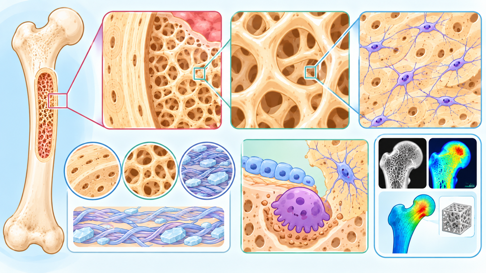
骨骼并不是一块均匀、坚硬的“钙块”,而是一个具有高度层级化结构的生物复合材料。从宏观的股骨、椎体,到毫米尺度的皮质骨和松质骨,再到微米尺度的骨小梁、骨陷窝—骨小管网络以及胶原—矿物质复合结构,骨的强度来自这些不同尺度结构之间的协同作用。因此,骨密度(BMD)只是骨骼健康的一部分,骨微结构(bone microarchitecture)同样决定骨骼能否承受机械负荷以及发生骨折的可能性。近年来的研究越来越强调,两个BMD相似的人,其骨折风险仍可能明显不同,原因之一正是骨微结构存在差异
Bone is not simply a homogeneous block of mineral. It is a highly organized biological composite material extending across multiple structural scales—from the whole femur or vertebral body to cortical and trabecular compartments, and further down to trabeculae, osteocyte lacunae-canalicular networks, and collagen–mineral composites. Skeletal strength emerges from the interaction of these structures. Therefore, bone mineral density (BMD) represents only one aspect of skeletal health; bone microarchitecture is also critical in determining mechanical competence and fracture susceptibility. Increasing evidence shows that people with similar BMD values can have substantially different fracture risks, partly because their underlying bone microarchitecture differs.
1. 皮质骨与松质骨:骨骼的两种基本建筑形式
骨微结构首先可以从两个主要组成部分理解:皮质骨(cortical bone)和松质骨(trabecular bone)。皮质骨构成骨骼外层坚硬的“壳”,在长骨骨干尤其发达,能够抵抗弯曲和扭转。松质骨则由相互连接的骨小梁组成,主要存在于椎体、股骨近端和长骨末端等区域。骨小梁并不是随机排列的,而是根据长期受到的机械载荷形成具有方向性的三维网络。骨骼因此能够以相对较低的质量获得较高的力学效率。
Bone microarchitecture can first be understood through its two major compartments: cortical bone and trabecular bone. Cortical bone forms the dense outer shell of the skeleton and is particularly prominent in the shafts of long bones, where it resists bending and torsion. Trabecular bone consists of an interconnected three-dimensional network of trabeculae and is abundant in vertebrae, the proximal femur, and the ends of long bones. Trabeculae are not randomly organized; their orientation and connectivity adapt to habitual mechanical loading, allowing the skeleton to achieve considerable mechanical efficiency with relatively little mass.
2. 骨小梁:并不是“越多越好”
评价松质骨微结构时,研究人员通常关注骨小梁体积分数(BV/TV)、骨小梁厚度(Tb.Th)、数量(Tb.N)、间距(Tb.Sp)以及连接性和各向异性等参数。健康骨骼需要在这些指标之间保持合理的组合。骨质疏松并不只是“骨小梁变少”,还可能表现为骨小梁变薄、断裂、连接减少以及原本的板状结构逐渐转化为更脆弱的杆状结构。这样的变化会显著削弱三维骨架的承载能力,即使总体BMD下降并不十分明显,也可能增加骨折风险。
Trabecular microarchitecture is commonly characterized by parameters such as bone volume fraction (BV/TV), trabecular thickness (Tb.Th), trabecular number (Tb.N), trabecular separation (Tb.Sp), connectivity, and anisotropy. Healthy bone requires an appropriate combination of these properties. Osteoporosis is not simply a matter of “fewer trabeculae.” Trabeculae may become thinner, disconnected, and structurally degraded, with plate-like structures becoming more rod-like and mechanically vulnerable. These alterations can substantially weaken the three-dimensional load-bearing network, sometimes increasing fracture risk even when the reduction in overall BMD is relatively modest.
3. 皮质骨:厚度之外,还有一个关键指标——孔隙率
皮质骨同样具有复杂的微结构。随着年龄增长以及绝经后雌激素水平下降,皮质骨可能逐渐变薄,同时出现皮质骨孔隙率(cortical porosity)增加。这些微小孔道与血管、骨髓以及骨重塑单位有关。当孔隙数量和尺寸增加时,皮质骨的有效承载截面下降,局部应力集中增加,最终可能明显降低骨骼的抗骨折能力。因此,骨折风险不能简单地用“皮质骨厚不厚”来解释,还需要考虑皮质骨内部的孔隙结构和空间分布。
Cortical bone also possesses a sophisticated microstructure. With ageing and declining estrogen levels after menopause, cortical bone can become thinner while cortical porosity increases. These microscopic pores are associated with vascular channels, marrow spaces, and remodeling activity. As the number and size of pores increase, the effective load-bearing area of the cortex decreases and local stress concentrations may rise. Consequently, cortical fracture resistance can deteriorate substantially. Cortical thickness alone is therefore insufficient; the size, distribution, and organization of cortical pores are also important determinants of skeletal strength.
4. 骨细胞:微结构中的“传感器”
在骨组织内部,还存在一种极其特殊的细胞——骨细胞(osteocyte)。成熟骨细胞被包埋在骨基质中的骨陷窝内,并通过细长的细胞突起形成复杂的骨小管网络。这个网络不仅负责细胞之间的信息交流,也是骨骼感知机械负荷的重要结构。2026年的多尺度建模研究进一步表明,骨细胞骨陷窝本身会改变局部应力和应变分布,可能影响机械信号传递以及微损伤的起始位置。这意味着,骨微结构不仅是“承重结构”,还是一个能够参与**机械感知(mechanosensing)**的动态生物系统。
Deep within the bone matrix lies a highly specialized cell: the osteocyte. Mature osteocytes reside in microscopic lacunae and extend long cellular processes through the canalicular network. This system enables communication between osteocytes and provides a major structural basis for skeletal mechanosensing. A 2026 multiscale modeling study showed that osteocyte lacunae themselves perturb local stress and strain fields and may influence both mechanotransduction and the initiation of microdamage. Bone microarchitecture is therefore not merely a passive load-bearing framework; it is also an active biological system involved in mechanical sensing and adaptation.
5. 微损伤:骨骼为什么需要不断重塑?
日常生活中,骨骼不断受到走路、跑步、跳跃以及肌肉收缩产生的机械应力。即使没有发生明显骨折,骨组织内部也会产生微小损伤。健康骨骼能够通过骨重塑不断识别并清除这些区域,再形成新的骨组织。如果微损伤积累速度超过修复能力,就可能造成局部结构薄弱。因此,骨重塑实际上是在不断维护骨微结构的“安全性”。这也解释了为什么骨量相同的两块骨,其力学性能可能完全不同。
During everyday activities, bone is continuously exposed to mechanical forces generated by walking, running, jumping, and muscle contraction. Even in the absence of an obvious fracture, microscopic damage can accumulate within the bone matrix. Healthy bone continuously detects and repairs such damage through remodeling. When microdamage accumulates faster than it can be repaired, localized structural weakness may develop. Bone remodeling can therefore be viewed as a continuous maintenance system that preserves the structural safety of bone microarchitecture. This helps explain why two bones with similar amounts of mineral may nevertheless have very different mechanical properties.
6. 为什么BMD不能完全代表骨强度?
DXA测量的骨密度主要反映单位面积上的骨矿物质含量,是目前临床诊断骨质疏松的重要基础指标。然而,BMD并不能直接告诉我们矿物质究竟如何分布,也不能完整描述骨小梁连接性、皮质骨孔隙率、胶原性质和局部应力分布。2025年的综述指出,骨骼实际上是由矿物质、胶原基质和水共同构成的复合材料,而其复杂的层级结构决定了骨骼同时具有刚性、韧性以及耗散能量的能力。
DXA-derived BMD primarily reflects areal bone mineral content and remains a fundamental clinical measure for osteoporosis. However, BMD does not reveal how mineral is distributed within bone, nor does it fully capture trabecular connectivity, cortical porosity, collagen properties, or local stress concentrations. A 2025 review emphasized that bone is a composite of mineral, collagen matrix, and water, organized into a hierarchical structure that allows it to combine stiffness, toughness, and energy dissipation.
7. HR-pQCT:让骨微结构“看得见”
近年来,高分辨率外周定量计算机断层扫描(HR-pQCT)成为研究骨微结构的重要工具。与传统DXA的二维投影不同,HR-pQCT能够获得接近三维的高分辨率骨结构信息,并分别分析皮质骨和松质骨。2025年的纵向研究显示,HR-pQCT获得的微结构和骨强度指标能够帮助预测长期糖皮质激素使用者的脆性骨折风险,而这种风险并不能完全由传统BMD或FRAX解释。
High-resolution peripheral quantitative computed tomography (HR-pQCT) has become an important research technology for visualizing bone microarchitecture. Unlike the two-dimensional projection obtained by DXA, HR-pQCT provides high-resolution three-dimensional information and allows separate assessment of cortical and trabecular compartments. A 2025 longitudinal study found that HR-pQCT-derived microstructural and estimated strength parameters could improve prediction of fragility fractures in patients receiving long-term glucocorticoids, suggesting information beyond that provided by conventional BMD or FRAX.
8. 2026年的新技术:Photon-counting CT
骨微结构成像正在进入新的阶段。**光子计数CT(photon-counting CT,PCCT)**能够直接利用单个X射线光子的能量信息,相较传统能量积分型CT具有更高的空间和对比度分辨能力,并有潜力进行材料分解。2026年的研究表明,PCCT在临床可接受的辐射剂量下,可以获得与HR-pQCT高度相关的骨小梁结构参数;最新综述则认为,PCCT未来有可能同时用于骨微结构和骨矿物质密度评价。不过,目前仍需要解决辐射剂量、重建算法以及真实临床环境下标准化等问题。
Bone microarchitecture imaging is now entering a new technological phase. Photon-counting CT (PCCT) detects individual X-ray photons and their energy information, offering improved spatial and contrast resolution compared with conventional energy-integrating CT and enabling material decomposition. Studies published in 2026 have demonstrated strong agreement between PCCT and HR-pQCT for trabecular microstructural measurements at clinically acceptable radiation doses. Recent reviews suggest that PCCT may eventually provide simultaneous assessment of bone microarchitecture and mineral density, although radiation dose, reconstruction algorithms, and clinical standardization remain important challenges.
9. 从二维骨密度到三维“骨骼地图”
另一个值得关注的方向是3D-DXA(三维双能X线吸收测定)。它可以利用常规DXA数据重建近端股骨的三维模型,并估算皮质和松质骨相关参数。2026年一项涵盖531人的多队列研究发现,3D-DXA得到的多个参数与QCT测量结果具有较强相关性,相关系数达到0.82–0.97。这意味着未来部分骨微结构信息可能不一定需要额外进行CT扫描,而可以从常规DXA中获得更多三维信息。
Another emerging approach is 3D-DXA, which reconstructs three-dimensional models of the proximal femur from standard DXA scans and estimates cortical and trabecular parameters. A 2026 multicohort study involving 531 participants found strong correlations between 3D-DXA measurements and QCT-derived parameters, with correlation coefficients ranging from 0.82 to 0.97. This raises the possibility that future bone assessment could extract substantially more three-dimensional structural information from routine DXA examinations without requiring an additional CT scan.
10. AI正在改变我们理解骨微结构的方式
人工智能可能进一步改变骨微结构评估。传统方法往往需要人工设定区域、提取结构参数,再根据统计模型评估骨折风险;而AI可以同时处理影像中的骨密度、形态、空间结构甚至纹理信息。2026年的综述指出,AI辅助的常规影像分析、TBS、3D形态建模以及有限元分析等方法正在逐渐形成传统BMD之外的补充性骨质量评估体系。未来真正有价值的AI模型,可能不只是告诉医生“BMD是多少”,而是进一步回答:骨骼哪里最脆弱、为什么脆弱,以及未来发生骨折的风险在哪里。
Artificial intelligence may further transform the assessment of bone microarchitecture. Traditional analysis often requires predefined regions, extraction of structural parameters, and statistical modeling of fracture risk. AI can potentially integrate density, morphology, spatial organization, and image texture simultaneously. A 2026 review highlighted the growing role of AI-assisted routine imaging analysis, trabecular bone score, three-dimensional shape modeling, and finite-element analysis as complementary approaches to conventional BMD. The ultimate goal may not simply be to report “What is the BMD?”, but rather to determine where the skeleton is structurally vulnerable, why it is vulnerable, and where fracture risk is concentrated.
结语|Conclusion
骨微结构是理解骨骼强度和骨折风险的关键。骨骼真正的“力量”并不只来自其中含有多少钙,而来自皮质骨厚度、皮质骨孔隙率、骨小梁数量和连接性、胶原与矿物质组成,以及骨细胞所构成的机械感知网络。骨质疏松也因此不是单纯的“骨量减少”,而是一个涉及结构、材料和生物学功能的综合性疾病。
Bone microarchitecture is fundamental to understanding skeletal strength and fracture risk. The true “strength” of bone depends not simply on how much calcium it contains, but on the integrated properties of cortical thickness, cortical porosity, trabecular number and connectivity, collagen–mineral composition, and the mechanosensory network formed by osteocytes. Osteoporosis should therefore be understood not merely as a loss of bone quantity, but as a disease involving structural, material, and biological deterioration.
未来,骨健康评价可能逐渐从单一的BMD指标走向**“Bone Quality + Bone Microarchitecture + Bone Strength + Dynamic Remodeling”的综合评价体系。HR-pQCT、PCCT、3D-DXA、先进MRI、有限元分析以及人工智能正在把过去无法直接观察的骨微结构逐渐变成可以量化、建模和预测的数据。对骨骼而言,这意味着我们正在从“测量有多少骨”,走向真正理解“骨是怎样组织起来的,以及它为什么会断”**。
In the future, skeletal assessment may increasingly evolve from a single BMD measurement toward an integrated framework of Bone Quality + Bone Microarchitecture + Bone Strength + Dynamic Remodeling. HR-pQCT, PCCT, 3D-DXA, advanced MRI, finite-element modeling, and artificial intelligence are progressively transforming previously inaccessible aspects of bone microstructure into measurable and predictive information. In other words, bone science is moving from simply asking “How much bone is there?” toward understanding “How is the bone organized, and why does it fracture?”
参考文献|References
- Patzer TS, Schwartz FR, Panta RK, et al. Assessing bone microstructure and density with photon-counting CT: emerging applications and challenges. Skeletal Radiology. 2026;55:2161–2174.
- Peña JA, Thomsen F, Damm T, et al. Assessment of trabecular microstructure using photon-counting CT: Influence of dose, spatial resolution and reconstruction algorithms. Bone. 2026;213:118066.
- Bracco MI, Black DM, Sone T, et al. Validation of 3D-DXA-derived proximal femur measurements against QCT across international clinical cohorts. Bone. 2026;211:117988.
- Clementini L, Vergani LM, Buccino F. Image-guided multiscale modeling of trabecular bone: lacuna-resolved stress amplification and interaction effects. Acta Biomaterialia. 2026.
- Zhang H, Xiao P, Ye K, Wang X. Conformity to stochastic invariance of microstructure influences mechanical competence of trabecular bone. Bone. 2026;209:117925.
- Liu Z, Li S, Silberschmidt VV. New local trabecular support ranks formation-labelled transitions: An image-derived framework for bone adaptation in human distal-tibia based on HR-pQCT. Bone. 2026;213:118052
- So H, Griffith JF, Lau SL, et al. Bone microarchitecture assessed by 3D high-resolution peripheral quantitative computed tomography as predictor of fracture risk in patients with glucocorticoid-induced osteoporosis. Journal of Orthopaedic Translation. 2025;51:329–336.
- Shin SH, Chae HD, Suprana A, et al. UTE MRI technical developments and applications in osteoporosis: a review. Frontiers in Endocrinology. 2025;16:1510010.
- Single-component imaging and ultrasound-based tools for the assessment of fracture risk. Nature Reviews Rheumatology. 2026.
- Two Decades of High-Resolution Peripheral Quantitative Computed Tomography: Present and Future Clinical Perspectives. 2024.