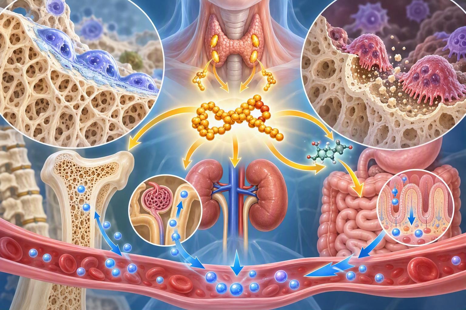
甲状旁腺激素(parathyroid hormone,PTH)是人体钙磷代谢和骨代谢中最重要的激素之一。它由甲状旁腺主细胞分泌,主要作用是帮助机体维持血钙稳定。当血液中的离子钙下降时,甲状旁腺会增加 PTH 分泌,促进肾脏减少钙排泄、促进活性维生素D生成,并通过作用于骨组织调节钙的释放。因此,PTH 并不仅仅是一种“调节骨骼的激素”,而是连接骨、肾脏和肠道钙磷代谢的重要内分泌信号。
Parathyroid hormone (PTH) is one of the central hormones regulating calcium and phosphate homeostasis and skeletal metabolism. It is secreted by the chief cells of the parathyroid glands and plays a major role in maintaining stable circulating calcium concentrations. When ionized calcium falls, PTH secretion increases, promoting renal calcium retention, stimulating the production of active vitamin D, and altering mineral flux between bone and the circulation. PTH therefore functions not simply as a “bone hormone,” but as an endocrine signal connecting the skeleton, kidneys, and intestinal mineral metabolism.
1. PTH如何感知血钙变化?
PTH 的分泌受到**钙敏感受体(calcium-sensing receptor,CaSR)**精细调节。当血钙下降时,CaSR 所接受的抑制信号减弱,甲状旁腺细胞释放更多 PTH;当血钙恢复时,PTH 分泌则受到抑制。这种负反馈机制使血钙维持在一个相对狭窄的生理范围内。PTH 同时作用于肾脏,增加远端肾小管钙重吸收,并促进肾脏将 25-羟维生素D转化为具有生物活性的 1,25-二羟维生素D,也就是骨化三醇。
PTH secretion is tightly controlled by the calcium-sensing receptor (CaSR). When extracellular calcium decreases, inhibitory signaling through CaSR is reduced and parathyroid cells release more PTH. As calcium levels recover, PTH secretion is suppressed. This negative-feedback system maintains circulating calcium within a narrow physiological range. PTH also acts on the kidney to increase calcium reabsorption and stimulate conversion of 25-hydroxyvitamin D into the biologically active hormone 1,25-dihydroxyvitamin D, or calcitriol.
2. PTH与骨组织之间的关键连接点:PTH1R
骨细胞本身并不是简单地被 PTH “直接命令”。PTH 主要通过**甲状旁腺激素1型受体(PTH1R)**发挥作用。PTH1R 广泛存在于成骨细胞谱系细胞和骨细胞中。当 PTH 与 PTH1R 结合后,可以激活 G 蛋白偶联信号通路,包括 cAMP/PKA 和其他下游信号网络,从而改变成骨细胞、骨细胞以及骨重塑相关细胞的行为。
The major molecular connection between PTH and bone is the parathyroid hormone 1 receptor (PTH1R). PTH1R is expressed predominantly by osteoblast-lineage cells and osteocytes. Binding of PTH to PTH1R activates G-protein-coupled signaling pathways, including cAMP/PKA and several downstream networks. These signals alter the behavior of osteoblasts, osteocytes, and other cells involved in bone remodeling.
3. 为什么PTH有时“增加骨吸收”,有时却“增加骨形成”?
这是理解 PTH 与骨代谢关系的核心。持续升高的 PTH,例如原发性甲状旁腺功能亢进时,可以促进骨转换,并最终使骨吸收占据优势;但短暂、间歇性地刺激 PTH1R,却能够产生明显的促骨形成效应。也就是说,同一种激素、同一个受体,由于刺激的时间模式不同,可以产生截然不同的骨代谢结果。这种“时间编码”现象是内分泌信号研究中的一个重要概念。
This apparent paradox is central to understanding PTH biology. Chronically elevated PTH, as occurs in primary hyperparathyroidism, can increase bone turnover and favor net bone loss in susceptible skeletal compartments. In contrast, short, intermittent stimulation of PTH1R produces a net anabolic effect on bone. Thus, the same hormone acting through the same receptor can generate very different skeletal outcomes depending on the temporal pattern of receptor stimulation. This concept of “temporal encoding” is a fundamental feature of PTH signaling.
4. 间歇性PTH为什么能够促进骨形成?
间歇性 PTH 刺激可以迅速改变成骨细胞谱系的基因表达和细胞行为。研究显示,PTH 能够促进成骨细胞存活、增加成骨相关活性,并影响 Wnt/β-catenin 等骨形成信号通路。与此同时,PTH 还能够改变骨细胞产生的 Sclerostin 等信号分子的水平,从而解除部分对 Wnt 通路的抑制。最终结果是,新的骨形成在治疗早期超过骨吸收,形成所谓的 anabolic window(合成代谢窗口)。
Intermittent PTH exposure rapidly changes gene expression and cellular behavior in the osteoblast lineage. PTH can enhance osteoblast survival and activity and influence osteogenic pathways including Wnt/β-catenin signaling. It can also alter osteocyte-derived signals such as sclerostin, thereby reducing inhibition of Wnt-mediated bone formation. During the early phase of treatment, bone formation can therefore exceed resorption, creating an anabolic window in which net bone mass increases.
5. PTH其实也会促进破骨细胞
一个容易产生误解的地方是:PTH 并不是只“刺激成骨细胞”。成骨细胞谱系细胞和骨细胞可以产生 RANKL,而 PTH 能够影响 RANKL/OPG 平衡,从而促进破骨细胞生成。因此,PTH 在一定条件下同时增加骨形成和骨吸收。间歇性给药之所以最终能够增加骨量,并不是因为破骨细胞完全不受影响,而是因为早期的成骨反应在特定时间窗口内超过了骨吸收。
An important nuance is that PTH does not exclusively stimulate osteoblasts. Osteoblast-lineage cells and osteocytes can produce RANKL, and PTH can alter the RANKL/OPG balance, thereby promoting osteoclastogenesis. Consequently, intermittent PTH treatment can increase both bone formation and resorption. The net anabolic response does not result from completely suppressing osteoclasts; rather, during the early treatment window, the increase in bone formation exceeds the accompanying increase in resorption.
6. 最新研究正在重新定义“PTH促进骨形成”的机制
2025 年发表的组织形态学综述对人体间歇性 PTH 治疗后的骨重塑进行了重新分析。研究者发现,治疗最初约 6 个月时,成骨细胞和破骨细胞的活性都会增加;但骨形成的增加并不能简单归结为传统意义上的“建模性骨形成”。研究提示,**由骨吸收启动的重塑性骨形成(remodeling-based bone formation)**可能是 PTH 促骨形成的重要组成部分。这意味着 PTH 的作用实际上与完整的骨重塑耦联网络密切相关。
A 2025 histomorphometric review of intermittent PTH treatment in humans has refined this model. During approximately the first six months of therapy, both osteoblast and osteoclast activity increase. Importantly, the anabolic response cannot simply be explained by classical modeling-based bone formation. The evidence suggests that remodeling-based bone formation initiated by osteoclastic activity may represent a major component of the PTH response. This reinforces the concept that PTH-induced anabolism is closely linked to coordinated bone remodeling rather than isolated osteoblast activation.
7. Teriparatide:PTH进入骨质疏松治疗领域
这种机制最终被转化为临床治疗。**Teriparatide(特立帕肽)**是重组人 PTH 的 1–34 氨基酸片段,通过间歇性刺激 PTH1R 发挥促骨形成作用。它能够增加脊柱、髋部等部位的骨密度,并降低椎体和部分非椎体骨折风险。对于骨折风险非常高的骨质疏松患者,PTH1R 激动剂属于重要的骨形成治疗策略。
This biology has been translated into clinical medicine. Teriparatide, a recombinant fragment corresponding to PTH(1–34), produces an anabolic skeletal response through intermittent PTH1R stimulation. It increases bone mineral density at sites including the lumbar spine and hip and reduces vertebral and non-vertebral fracture risk. PTH1R agonists therefore represent an important class of osteoanabolic therapy for patients at high risk of osteoporotic fracture.
8. Abaloparatide:另一种PTH1R激动剂
另一种重要药物是 Abaloparatide(阿巴洛帕肽)。它与 PTH 相关蛋白(PTHrP)具有结构和功能上的联系,同样通过 PTH1R 发挥作用。2025 年发表的系统综述和网络荟萃分析显示,特立帕肽和阿巴洛帕肽均能降低椎体及非椎体骨折风险;部分分析还提示阿巴洛帕肽在非椎体骨折方面可能具有一定优势。不过,这些比较需要结合研究设计、患者特征和治疗方案进行解释,不能简单理解为一种药物全面优于另一种。
Another important PTH1R agonist is abaloparatide, which is structurally related to parathyroid hormone-related protein (PTHrP). A 2025 systematic review and network meta-analysis found that both teriparatide and abaloparatide reduce vertebral and non-vertebral fracture risk. Some analyses suggested a potential advantage of abaloparatide for non-vertebral fracture outcomes, although such comparisons must be interpreted in the context of study populations, trial design, and treatment sequences rather than as evidence that one drug is universally superior.
9. 一个新的问题:为什么PTH的促骨作用会逐渐减弱?
近年来一个非常重要的问题是:为什么长期使用 PTH 类药物后,骨形成效应会逐渐减弱? 2026 年发表的研究综述指出,PTH 促骨形成作用通常在治疗早期最明显,随后逐渐减弱。可能机制包括成骨祖细胞数量或活性的限制、PTH1R 信号适应性变化、骨形成表面逐渐饱和,以及 Sclerostin、DKK1 等 Wnt 抑制因子的反馈调节。
A major current research question is: why does the anabolic effect of PTH gradually diminish with prolonged treatment? A 2026 review highlighted several possible mechanisms, including limitations in osteoprogenitor availability, adaptive changes in PTH1R signaling, saturation of bone-forming surfaces, and increased counter-regulatory activity involving Wnt inhibitors such as sclerostin and DKK1. The decline in response appears to be a biological limitation of the remodeling system rather than simply a pharmacological failure.
10. 2026年的研究:MCP-1可能参与PTH的促骨作用
2026 年的动物研究还进一步关注 MCP-1/CCL2。研究发现,PTH 给药后骨组织中的 MCP-1 表达可以迅速升高;在特定遗传模型中,如果成骨细胞/骨细胞谱系缺失 MCP-1,PTH 的促骨形成效应会受到明显影响。这一结果提示,趋化因子并不仅仅参与免疫反应,也可能参与 PTH 调节骨形成的细胞通讯过程。不过,目前这些证据主要来自实验动物,距离临床治疗应用仍有明显距离。
A 2026 experimental study has also focused on MCP-1/CCL2 as a possible mediator of PTH-induced anabolism. PTH rapidly increased MCP-1 expression in bone, and selective deletion of MCP-1 in the osteoblast/osteocyte lineage impaired the anabolic response to PTH in experimental models. These findings suggest that chemokines may participate in the cellular communication underlying PTH-mediated bone formation. However, this evidence remains preclinical and should not yet be interpreted as a validated therapeutic target in humans.
11. PTH与甲状旁腺功能亢进:同一种激素的另一面
如果 PTH 长期处于异常升高状态,情况就完全不同。例如原发性甲状旁腺功能亢进可导致持续性的骨转换增加,并可能造成骨量下降。慢性肾脏疾病患者还可能出现继发性甲状旁腺功能亢进,使 PTH、磷、钙和维生素D代谢发生复杂变化。因此,在临床上看到“PTH升高”时,不能简单地认为它意味着“骨形成增强”,而必须结合血钙、磷、25-羟维生素D、肾功能以及骨密度等指标进行综合判断。
When PTH remains chronically elevated, the biological outcome can be very different. Primary hyperparathyroidism can produce sustained high bone turnover and, in some patients, substantial skeletal deterioration. Patients with chronic kidney disease may develop secondary hyperparathyroidism, in which PTH interacts with abnormalities in phosphate, calcium, vitamin D, and mineral metabolism. Therefore, an elevated PTH concentration should never be interpreted simply as evidence of increased bone formation. Clinical interpretation requires integration of calcium, phosphate, vitamin D status, kidney function, and skeletal measurements.
12. PTH让我们重新认识骨代谢
PTH 最有趣的地方在于,它说明骨代谢并不是简单的“激素升高=骨吸收,激素降低=骨形成”。真正重要的是信号的强度、持续时间、频率以及细胞所处的生理环境。从传统的钙稳态激素,到今天的骨质疏松促骨形成药物,再到 2025–2026 年对 PTH1R、Wnt、RANKL/OPG、MCP-1 和骨重塑耦联机制的深入研究,PTH 已经成为理解骨代谢调控网络的重要窗口。
The biology of PTH demonstrates that bone metabolism cannot be reduced to a simple equation in which “high hormone means resorption” and “low hormone means formation.” The biological outcome depends on the intensity, duration, frequency, and cellular context of signaling. From a classical regulator of calcium homeostasis to an established osteoanabolic therapeutic pathway, and now to an active area of research involving PTH1R, Wnt signaling, RANKL/OPG, MCP-1, and remodeling coupling, PTH remains one of the most informative models for understanding how the skeleton is regulated.
结语|Conclusion
甲状旁腺激素连接着血钙稳态、肾脏功能和骨重塑,是人体矿物质代谢系统中的核心调节因子。它最令人着迷的地方在于:持续刺激可能促进骨吸收,而合理的间歇性刺激却能够促进骨形成。 这种看似矛盾的现象实际上揭示了细胞信号“时间模式”的重要性。未来研究的重点,很可能不再只是寻找更强的 PTH 类药物,而是寻找能够更精准控制 PTH1R 信号持续时间、细胞类型和下游通路的方法,从而延长促骨形成作用,并更好地维持骨重塑的长期平衡。
Parathyroid hormone links calcium homeostasis, kidney function, and skeletal remodeling and is therefore a central regulator of mineral metabolism. Its most remarkable feature is that chronic stimulation can favor bone resorption, whereas appropriately timed intermittent stimulation can promote bone formation. This apparent paradox reveals the importance of the temporal pattern of cellular signaling. Future research may increasingly focus not simply on developing stronger PTH analogues, but on precisely controlling the duration, cellular specificity, and downstream consequences of PTH1R signaling to extend anabolic activity while preserving long-term remodeling balance.
参考文献|References
- Fuggle N, Rizzoli R, Beaudart C, et al. Parathyroid hormone receptor agonists in the management of osteoporosis. Nature Reviews Rheumatology. 2025;21:599–611. DOI: 10.1038/s41584-025-01287-w.
- Beaudart C, Veronese N, Douxfils J, et al. PTH1 receptor agonists for fracture risk: a systematic review and network meta-analysis. Osteoporosis International. 2025;36(6):951–967. DOI: 10.1007/s00198-025-07440-1.
- Koch Thomsen L, van Dijk Christiansen P, Møller Andreasen C, Levin Andersen T. The cellular effect of intermittent PTH treatment on bone remodeling and modeling in humans—a histomorphometry centered scoping review. Osteoporosis International. 2025;36(12):2411–2435. DOI: 10.1007/s00198-025-07612-z.
- Mechanisms limiting the long-term anabolic effects of teriparatide (PTH 1-34) on bone. JBMR Plus. 2026;10(7):ziag093. Published May 2026.
- Mechanisms underlying the waning of osteoanabolic therapy effects in osteoporosis. 2026. Review of the mechanisms underlying attenuation of PTH analogue and other osteoanabolic therapies.
- Anabolic effect of parathyroid hormone (1-34) to prevent ovariectomy induced bone loss is attenuated in Col1A1-cre floxed monocyte chemotactic protein 1 (MCP1, CCL2) mice. Bone. 2026; DOI: 10.1016/j.bone.2026.117898.
- Jørgensen NR, Cavalier E, Vasikaran SD. Parathyroid hormone assays in 2026: second versus third generation, standardization, and clinical interpretation. Current Opinion in Endocrinology, Diabetes and Obesity. 2026;33(4):125–130. DOI: 10.1097/MED.0000000000000960.